Clinical Focus ›› 2025, Vol. 40 ›› Issue (12): 1147-1152.doi: 10.3969/j.issn.1004-583X.2025.12.017
-
Received:2025-11-05Online:2025-12-20Published:2025-12-30
CLC Number:
Cite this article
share this article
Add to citation manager EndNote|Ris|BibTeX
URL: https://www.lchc.cn/EN/10.3969/j.issn.1004-583X.2025.12.017
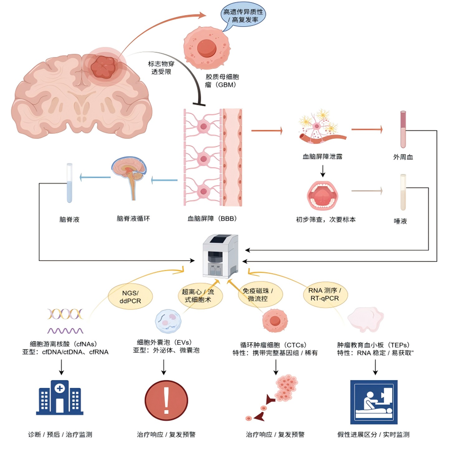
| 标志物类型 | 主要亚型 | 来源 | 检测技术 | 优势 | 局限性 | 临床应用方向 |
|---|---|---|---|---|---|---|
| 细胞游离DNA | cfDNA(其中肿瘤来源部分为ctDNA) | 肿瘤细胞坏死/凋亡 | NGS、ddPCR、PCR | 携带肿瘤突变/甲基化信息,可动态监测 | 外周血浓度低,受炎症干扰 | 诊断、预后评估、治疗响应监测 |
| 细胞游离RNA | cfRNA(miRNA、lncRNA) | 肿瘤细胞分泌 | RT-qPCR、NGS | 反映基因表达动态,组织特异性高 | 易降解,需EV保护或特殊保存 | 诊断、复发预警、治疗分层 |
| EVs | 外泌体(30~100 nm) | 肿瘤细胞内体途径 | 超离心、密度梯度离心、质谱 | 内容物稳定,可通过BBB | 分离无标准化protocol,检测成本高 | 诊断、治疗响应监测、靶向药物载体 |
| 微囊泡 | 微囊泡(0.1~1 μm) | 肿瘤细胞膜出芽 | 流式细胞术、免疫磁珠分选 | 易分离,蛋白标志物丰富 | 与其他囊泡区分困难 | 复发预警、炎症相关监测 |
| CTCs | GBM来源CTCs | 肿瘤原发灶脱落 | 免疫细胞化学、微流控芯片、FISH | 携带完整肿瘤基因组,反映侵袭能力 | 外周血稀有,检测敏感度低 | 分子分型、转移风险评估 |
| TEPs | TEPs | 血小板被肿瘤“教育” | RNA测序、RT-qPCR | RNA稳定性高,易获取 | 缺乏GBM特异性RNA标志物 | 诊断、假性进展区分、实时监测 |
| 生物标志物 | 检测样本 | 关键发现 | 检测技术 | 研究规模 (例) |
|---|---|---|---|---|
| ctDNA(TP53/EGFR) | 血浆 | 55%患者检测到突变,突变阳性者OS缩短3.2个月[ | NGS | 222 |
| cfDNA浓度 | 血浆 | 术前浓度>10 ng/ml者无进展生存期缩短4.5个月,OS缩短6.8个月[ | 荧光定量法 | 62 |
| EVs MGMT甲基化 | CSF | 预测替莫唑胺治疗响应的敏感度85.7%,特异度90%[ | ddPCR | 49 |
| 外泌体miR-19b-3p | 血清 | 诊断GBM的敏感度78%,特异度85%,与肿瘤增殖指数正相关[ | RT-qPCR | 87 |
| CTCs(GFAP+) | 外周血 | 放疗前检测率72%,放疗后降至8%,CTCs数量>3个/ml者复发风险升高3倍[ | 荧光报告系统 | 45 |
| TEPs RNA谱 | 全血 | 区分GBM与健康人的准确率95%,区分假性进展与真进展的准确率85%[ | RNA测序 | 120 |
| CSF miR-128/485-3p | CSF | 诊断GBM的敏感度82%,特异度91%,可区分WHO Ⅲ级与Ⅳ级胶质瘤[ | RT-qPCR | 63 |
| 微囊泡miR-625-5p | 血浆 | 术后表达降至正常,复发时升高2倍以上,预警复发的敏感度80%[ | RT-qPCR | 36 |
| [1] | King JL, Benhabbour SR. Glioblastoma multiforme-a look at the past and a glance at the future[J]. Pharmaceutics, 2021, 13(7): 1053. doi:10.3390/pharmaceutics13071053. |
| [2] | Ostrom QT, Cioffi G, Waite K, et al. CBTRUS statistical report: Primary brain and other central nervous system tumors diagnosed in the United States in 2014-2018[J]. Neuro Oncol, 2021, 23(Suppl 1): iii1-iii105. doi:10.1093/neuonc/noab001. |
| [3] |
Ohgaki H, Kleihues P. Genetic pathways to primary and secondary glioblastoma[J]. Am J Pathol, 2007, 170(5): 1445-1453. doi:10.1016/S0002-9440(10)63735-8.
pmid: 17456751 |
| [4] |
Ammirati M, Chotai S, Newton H, et al. Hypofractionated intensity modulated radiotherapy with temozolomide in newly diagnosed glioblastoma multiforme[J]. J Clin Neurosci, 2014, 21(4): 633-637. doi:10.1016/j.jocn.2013.10.018.
pmid: 24380758 |
| [5] |
Aldape K, Zadeh G, Mansouri S, et al. Glioblastoma: Pathology, molecular mechanisms and markers[J]. Acta Neuropathol, 2015, 129(6): 829-848. doi:10.1007/s00401-015-1448-4.
pmid: 25943888 |
| [6] |
Mannas JP, Lightner DD, Defrates SR, et al. Long-term treatment with temozolomide in malignant glioma[J]. J Clin Neurosci, 2014, 21(1): 121-123. doi:10.1016/j.jocn.2013.07.018.
pmid: 24063865 |
| [7] | Li Y, Ma Y, Wu Z, et al. Advanced imaging techniques for differentiating pseudoprogression and tumor recurrence after immunotherapy for glioblastoma[J]. Front Immunol, 2021, 12: 790674. doi:10.3389/fimmu.2021.790674. |
| [8] |
Garcia CM, Toms SA. The role of circulating microRNA in glioblastoma liquid biopsy[J]. World Neurosurg, 2020, 138: 425-435. doi:10.1016/j.wneu.2020.03.105.
pmid: 32251831 |
| [9] |
Chen M, Zhao H. Next-generation sequencing in liquid biopsy: Cancer screening and early detection[J]. Hum Genomics, 2019, 13(1): 34. doi:10.1186/s40246-019-0228-0.
pmid: 31370908 |
| [10] | Pantel K, Alix-Panabières C. Circulating tumour cells in cancer patients: Challenges and perspectives[J]. Trends Mol Med, 2010, 16(8): 398-406. doi:10.1016/j.molmed.2010.06.004. |
| [11] |
Pantel K, Alix-Panabières C. Liquid biopsy and minimal residual disease-latest advances and implications for cure[J]. Nat Rev Clin Oncol, 2019, 16(7): 409-424. doi:10.1038/s41571-019-0239-x.
pmid: 30796368 |
| [12] | Mandel P, Metais P. Nuclear acids in human blood plasma[J]. C R Seances Soc Biol Filiales, 1948, 142(3-4): 241-243. |
| [13] | Saenz-Antoñanzas A, Auzmendi-Iriarte J, Carrasco-Garcia E, et al. Liquid biopsy in glioblastoma: Opportunities, applications and challenges[J]. Cancers, 2019, 11(7): 950. doi:10.3390/cancers11070950. |
| [14] |
Bagley SJ, Till J, Abdalla A, et al. Clinical utility of plasma cell-free DNA in adult patients with newly diagnosed glioblastoma: A pilot prospective study[J]. Clin Cancer Res, 2020, 26(2): 397-407. doi:10.1158/1078-0432.CCR-19-2114.
pmid: 31666247 |
| [15] | Piccioni DE, Achrol AS, Kiedrowski LA, et al. Analysis of cell-free circulating tumor DNA in 419 patients with glioblastoma and other primary brain tumors[J]. CNS Oncol, 2019, 8(3): CNS34. doi:10.2217/cns-2019-0025. |
| [16] | Bagley SJ, Till J, Abdalla A, et al. Association of plasma cell-free DNA with survival in patients with IDH wild-type glioblastoma[J]. Neuro-Oncology Adv, 2021, 3(1): vdab011. doi:10.1093/noadv/vdab011. |
| [17] | Wang Y, Springer S, Zhang M, et al. Detection of tumor-derived DNA in cerebrospinal fluid of patients with primary tumors of the brain and spinal cord[J]. Proc Natl Acad Sci U S A, 2015, 112(32): 9704-9709. doi:10.1073/pnas.1509634112. |
| [18] | Mouliere F, Smith CG, Heider K, et al. Fragmentation patterns and personalized sequencing of cell-free DNA in urine and plasma of glioma patients[J]. EMBO Mol Med, 2021, 13(7): e12881. doi:10.15252/emmm.202012881. |
| [19] | Tomei S, Volontè A, Ravindran S, et al. MicroRNA expression profile distinguishes glioblastoma stem cells from differentiated tumor cells[J]. J Pers Med, 2021, 11(3): 264. doi:10.3390/jpm11030264. |
| [20] | Fontanilles M, Marguet F, Beaussire L, et al. Cell-free DNA and circulating TERT promoter mutation for disease monitoring in newly-diagnosed glioblastoma[J]. Acta Neuropathol Commun, 2020, 8(1): 179. doi:10.1186/s40478-020-01051-8. |
| [21] | Zan XY, Li L. Construction of lncRNA-mediated ceRNA network to reveal clinically relevant lncRNA biomarkers in glioblastomas[J]. Oncol Lett, 2019, 17(4): 4369-4374. doi:10.3892/ol.2019.10180. |
| [22] | Gao F, Cui Y, Jiang H, et al. Circulating tumor cell is a common property of brain glioma and promotes the monitoring system[J]. Oncotarget, 2016, 7(48): 71330-71340. doi:10.18632/oncotarget.12266. |
| [23] | Kopkova A, Sana J, Machackova T, et al. Cerebrospinal fluid microRNA signatures as diagnostic biomarkers in brain tumors[J]. Cancers, 2019, 11(10): 1546. doi:10.3390/cancers11101546. |
| [24] | Shen J, Hodges TR, Song R, et al. Serum HOTAIR and GAS5 levels as predictors of survival in patients with glioblastoma[J]. Mol Carcinog, 2018, 57(2): 137-141. doi:10.1002/mc.22744. |
| [25] | Zhang JX, Han L, Bao ZS, et al. HOTAIR, a cell cycle-associated long noncoding RNA and a strong predictor of survival, is preferentially expressed in classical and mesenchymal glioma[J]. Neuro-Oncology, 2013, 15(12): 1595-1603. doi:10.1093/neuonc/not184. |
| [26] | Wang Q, Song Y, Zhou C, et al. Combined detection of serum HOTAIR/GAS5 ratio predicts radiotherapy response and prognosis in glioblastoma[J]. Mol Ther Nucleic Acids, 2023, 35: 100-112. doi:10.1016/j.omtn.2023.05.012. |
| [27] | Cordonnier M, Chanteloup G, Isambert N, et al. Exosomes in cancer theranostic: Diamonds in the rough[J]. Cell Adh Migr, 2017, 11(2): 151-163. doi:10.1080/19336918.2017.1281943. |
| [28] |
Mondial A, Kumari Singh D, Panda S, et al. Extracellular vesicles as modulators of tumor microenvironment and disease progression in glioma[J]. Front Oncol, 2017, 7: 144. doi:10.3389/fonc.2017.00144.
pmid: 28730141 |
| [29] | Yang Q, Wei B, Peng C, et al. Identification of serum exosomal miR-98-5p, miR-183-5p, miR-323-3p and miR-19b-3p as potential biomarkers for glioblastoma patients and investigation of their mechanisms[J]. Curr Res Transl Med, 2022, 70: 103315. doi:10.1016/j.crm.2022.103315. |
| [30] |
Rosas-Alonso R, Colmenarejo-Fernández J, Pernía O, et al. Evaluation of the clinical use of MGMT methylation in extracellular vesicle-based liquid biopsy as a tool for glioblastoma patient management[J]. Sci Rep, 2024, 14(1): 11398. doi:10.1038/s41598-024-51696-4.
pmid: 38762534 |
| [31] | Döring K, Malinova V, Bettag C, et al. The diagnostic potential of extracellular vesicles derived from the blood plasma of glioblastoma patients[J]. In Vivo, 2024, 38(2): 2735-2739. doi:10.21873/invivo.134664. |
| [32] | Cilibrasi C, Simon T, Vintu M, et al. Definition of an inflammatory biomarker signature in plasma-derived extracellular vesicles of glioblastoma patients[J]. Biomedicines, 2022, 10(1): 125. doi:10.3390/biomedicines10010125. |
| [33] | Simionescu N, Nemecz M, Petrovici AR, et al. Microvesicles and microvesicle-associated microRNAs reflect glioblastoma regression: Microvesicle-associated miR-625-5p has biomarker potential[J]. Int J Mol Sci, 2022, 23(15): 8398. doi:10.3390/ijms23158398. |
| [34] |
Koch CJ, Lustig RA, Yang XY, et al. Microvesicles as a biomarker for tumor progression versus treatment effect in radiation/temozolomide-treated glioblastoma patients[J]. Transl Oncol, 2014, 7(6): 752-758. doi:10.1016/j.tranon.2014.09.004.
pmid: 25500085 |
| [35] | Zhang H, Yuan F, Qi Y, et al. Circulating tumor cells for glioma[J]. Front Oncol, 2021, 11: 607150. doi:10.3389/fonc.2021.607150. |
| [36] |
Van Schaaijik B, Wickremesekera AC, Mantamadiotis T, et al. Circulating tumor stem cells and glioblastoma: A review[J]. J Clin Neurosci, 2019, 61: 5-9. doi:10.1016/j.jocn.2018.10.024.
pmid: 30622004 |
| [37] | MacArthur KM, Kao GD, Chandrasekaran S, et al. Detection of brain tumor cells in the peripheral blood by a telomerase promoter-based assay[J]. Cancer Res, 2014, 74(7): 2152-2159. doi:10.1158/0008-5472.CAN-13-2861. |
| [38] |
Sullivan JP, Nahed BV, Madden MW, et al. Brain tumor cells in circulation are enriched for mesenchymal gene expression[J]. Cancer Discov, 2014, 4(11): 1299-1309. doi:10.1158/2159-8290.CD-14-0580.
pmid: 25139148 |
| [39] | Lynch D, Powter B, Po JW, et al. Isolation of circulating tumor cells from glioblastoma patients by direct immunomagnetic targeting[J]. Appl Sci, 2020, 10(10): 3338. doi:10.3390/app10103338. |
| [40] | Kichkailo AS, Narodov AA, Komarova MA, et al. Development of DNA aptamers for visualization of glial brain tumors and detection of circulating tumor cells[J]. Mol Ther Nucleic Acids, 2023, 32: 267-288. doi:10.1016/j.omtn.2023.01.020. |
| [41] |
Joosse SA, Pantel K. Tumor-educated platelets as liquid biopsy in cancer patients[J]. Cancer Cell, 2015, 28(5): 552-554. doi:10.1016/j.ccell.2015.10.004.
pmid: 26555171 |
| [42] | Best MG, Wesseling P, Wurdinger T. Tumor-educated platelets as a noninvasive biomarker source for cancer detection and progression monitoring[J]. Cancer Res, 2018, 78(12): 3407-3412. doi:10.1158/0008-5472.CAN-17-3416. |
| [43] | Sol N, et al. In ‘t Veld SGJG, Vancura A,Tumor-educated platelet RNA for the detection and (pseudo)progression monitoring of glioblastoma[J]. Cell Rep Med, 2020, 1(10): 100101. doi:10.1016/j.xcrm.2020.100101. |
| [44] |
Nilsson RJA, Balaj L, Hulleman E, et al. Blood platelets contain tumor-derived RNA biomarkers[J]. Blood, 2011, 118(13): 3680-3683. doi:10.1182/blood-2011-04-349848.
pmid: 21832279 |
| [45] | Aran V, De Melo Junior JO, Pilotto Heming C, et al. Unveiling the impact of corticosteroid therapy on liquid biopsy-detected cell-free DNA levels in meningioma and glioblastoma patients[J]. J Liquid Biopsy, 2024, 5(1): 100149. doi:10.1016/j.jlb.2024.100149. |
| [46] | Liu Y, Chen W, Zhang L, et al. Tumor in situ fluid circulating tumor DNA dynamically predicts recurrence and molecular residual disease in glioblastoma patients after surgery[J]. Neurosurgery, 2025, 97: 671-680. doi:10.1227/neu.0000000000002789. |
| [1] | Liu Fei, Lin Zhichun, Yue Jianlan, Yin Liang, Huang Shiming. Meta-analysis of different positron emission tomography/computed tomography imaging agents in the diagnosis of gliomas [J]. Clinical Focus, 2023, 38(4): 302-307. |
| [2] | Peng Yu, Jiang Zhihong, Wang Zhihai. Clinical and genetic analysis of a neonate with congenital central nervous system multiple malformation: An identification on mutations in VANGL1 genes [J]. Clinical Focus, 2022, 37(7): 640-643. |
| [3] | Zhang Zhaoming, Yu Xiangfeng, Song Jihui. Application value of diffusionweighted magnetic resonance imaging combined with spectral imaging in evaluating glioma grade [J]. Clinical Focus, 2020, 35(12): 1106-1110. |
| [4] | . [J]. Clinical Focus, 2016, 31(3): 337-339. |
| [5] | Li Xiaofu;Gao Ying;Han Zhongli. Evaluation of glioma tumor angiogenesis using enhanced T2 star weighted angiography [J]. Clinical Focus, 2015, 30(9): 1050-1053. |
| [6] | Liu Yang;Guo Qiang;Zhang Hao;Li Genhua;Feng Song;Jin Feng. Effect of Livin-targeted siRNA on production of TNF-αin TJ905 stem cells [J]. Clinical Focus, 2015, 30(3): 312-315. |
| [7] | . [J]. CLINICAL FOCUS, 2009, 24(9): 809-810. |
| Viewed | ||||||
|
Full text |
|
|||||
|
Abstract |
|
|||||