Clinical Focus ›› 2026, Vol. 41 ›› Issue (5): 411-416.doi: 10.3969/j.issn.1004-583X.2026.05.004
Previous Articles Next Articles
Artificial intelligence-assisted assessment of BI-RADS category 4 breast nodules by physicians of different seniority
Liu Xiaoli, Yang Shuang( ), Yin Li, Hu Yan, Zheng Yumeng, Duan Lihong
Department of Ultrasound ,Affiliated Zhongshan Hospital of Dalian University
-
Received:2025-10-15Online:2026-05-20Published:2026-05-26 -
Contact:Yang Shuang,Email: 1419861667@qq.com
CLC Number:
Cite this article
Liu Xiaoli, Yang Shuang, Yin Li, Hu Yan, Zheng Yumeng, Duan Lihong. Artificial intelligence-assisted assessment of BI-RADS category 4 breast nodules by physicians of different seniority[J]. Clinical Focus, 2026, 41(5): 411-416.
share this article
Add to citation manager EndNote|Ris|BibTeX
URL: https://www.lchc.cn/EN/10.3969/j.issn.1004-583X.2026.05.004
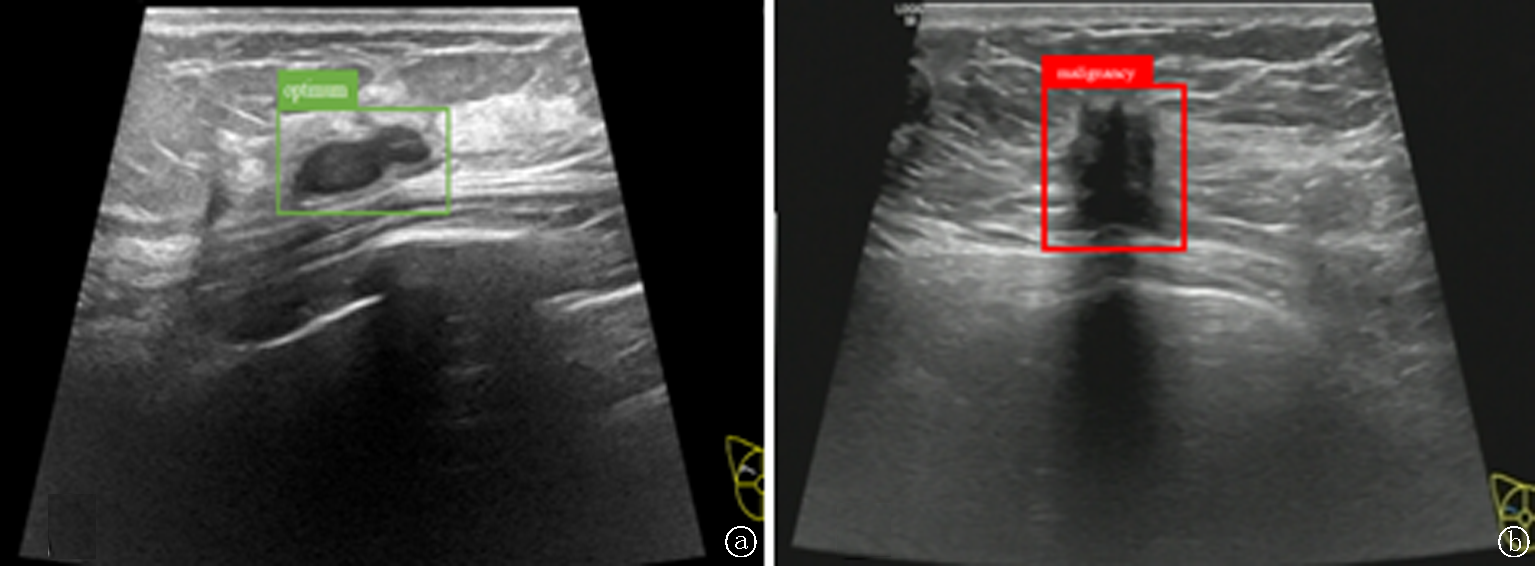
Fig.1 Imaging of Breast Nodules a. The YOLOv5 model suggested that the breast nodule was likely benign; histopathology confirmed fibroadenoma; b. The YOLOv5 model suggested that the breast nodule was likely malignant; histopathology confirmed invasive ductal carcinoma
| 诊断者 | 敏感度(%) | 特异度(%) | 准确度(%) | AUC(95%CI) |
|---|---|---|---|---|
| 低年资医师 | 64.91 | 65.38 | 65.18 | 0.618(0.520~0.716) |
| 中年资医师 | 80.70 | 74.35 | 77.03 | 0.767(0.683~0.850) |
| 高年资医师 | 92.98 | 83.33 | 87.40 | 0.863(0.795~0.932) |
| YOLOv5技术 | 73.68*#△ | 71.79* | 72.59*#△ | 0.733(0.648~0.819)*△ |
| 低年资+YOLOv5技术 | 80.70* | 70.51* | 74.81* | 0.782(0.705~0.859)* |
| 中年资+YOLOv5技术 | 91.22# | 73.07 | 80.74# | 0.872(0.810~0.933)# |
| 高年资+YOLOv5技术 | 91.22 | 80.76 | 85.18 | 0.878(0.818~0.938) |
Tab.1 Comparison of the diagnostic performance of physicians with different seniority levels, the YOLOv5 model, and combined diagnosis for BI-RADS category 4 breast nodules
| 诊断者 | 敏感度(%) | 特异度(%) | 准确度(%) | AUC(95%CI) |
|---|---|---|---|---|
| 低年资医师 | 64.91 | 65.38 | 65.18 | 0.618(0.520~0.716) |
| 中年资医师 | 80.70 | 74.35 | 77.03 | 0.767(0.683~0.850) |
| 高年资医师 | 92.98 | 83.33 | 87.40 | 0.863(0.795~0.932) |
| YOLOv5技术 | 73.68*#△ | 71.79* | 72.59*#△ | 0.733(0.648~0.819)*△ |
| 低年资+YOLOv5技术 | 80.70* | 70.51* | 74.81* | 0.782(0.705~0.859)* |
| 中年资+YOLOv5技术 | 91.22# | 73.07 | 80.74# | 0.872(0.810~0.933)# |
| 高年资+YOLOv5技术 | 91.22 | 80.76 | 85.18 | 0.878(0.818~0.938) |
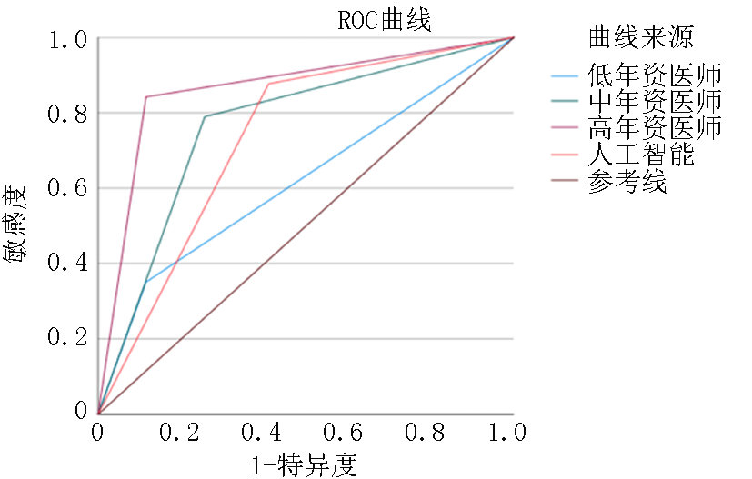
Fig.2 ROC curves of physicians with different seniority levels and the YOLOv5 model for predicting BI-RADS category 4 breast nodules
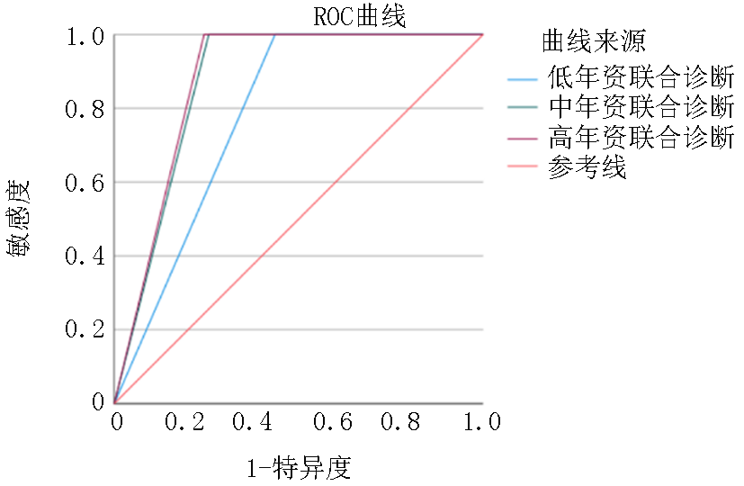
Fig.3 ROC curves of combined diagnosis by physicians with different seniority levels and the YOLOv5 model for predicting BI-RADS category 4 breast nodules
| 亚型 | 病理结果(例) | 低年资(%) | 中年资(%) | 高年资(%) | YOLOv5(%) | 低年资+AI(%) | 中年资+AI(%) | 高年资+AI(%) | ||
|---|---|---|---|---|---|---|---|---|---|---|
| 病灶数 | 恶性 | 良性 | ||||||||
| 4A | 76 | 17 | 59 | 59.21 | 73.68 | 84.21 | 69.74 | 71.05 | 82.89 | 84.21 |
| 4B | 41 | 24 | 17 | 70.73 | 82.93 | 87.80 | 73.17 | 82.93 | 87.80 | 87.80 |
| 4C | 18 | 16 | 2 | 83.33 | 88.89 | 94.44 | 83.33 | 88.89 | 94.44 | 94.44 |
Tab.2 Comparison of diagnostic accuracy of physicians with different seniority levels, the YOLOv5 model, and combined diagnosis for BI-RADS category 4 subtype nodules
| 亚型 | 病理结果(例) | 低年资(%) | 中年资(%) | 高年资(%) | YOLOv5(%) | 低年资+AI(%) | 中年资+AI(%) | 高年资+AI(%) | ||
|---|---|---|---|---|---|---|---|---|---|---|
| 病灶数 | 恶性 | 良性 | ||||||||
| 4A | 76 | 17 | 59 | 59.21 | 73.68 | 84.21 | 69.74 | 71.05 | 82.89 | 84.21 |
| 4B | 41 | 24 | 17 | 70.73 | 82.93 | 87.80 | 73.17 | 82.93 | 87.80 | 87.80 |
| 4C | 18 | 16 | 2 | 83.33 | 88.89 | 94.44 | 83.33 | 88.89 | 94.44 | 94.44 |
| [1] |
李玲玲, 苏荃利, 林菲菲, 等. 超声造影术前评价浸润性乳腺癌组织学分级的临床价值[J]. 中国超声医学杂志, 2022, 38(12):1350-1354. doi:10.3969/j.issn.1002-0101.2022.12.011.
|
| [2] |
吴墅, 赵佳琦, 陈蕊. 人工智能自动检测系统辅助超声诊断乳腺结节的临床应用[J]. 同济大学学报(医学版), 2022, 43(4):509-514. doi:10.12289/j.issn.1008-0392.22165.
|
| [3] |
金晓霞, 刘玉山, 陈鑫焱, 等. 三阴性乳腺癌中细胞分裂蛋白调节因子1的表达及新辅助化疗对其表达的影响[J]. 安徽医药, 2024, 28(12):2488-2491, 封3. doi:10.3969/j.issn.1009-6469.2024.12.031.
|
| [4] |
李文肖, 张玉瑞, 刘文, 等. S-Detect技术联合临床特征信息基于BI-RADS分类对乳腺肿块良恶性的诊断价值[J]. 现代肿瘤医学, 2025, 33(4):607-613. doi:10.3969/j.issn.1672-4992.2025.04.009.
|
| [5] |
刘文艳. 人工智能诊断系统在乳腺X线BI-RADS分类及乳腺良恶性病变鉴别诊断中的应用研究[D]. 苏州: 苏州大学, 2022.
|
| [6] |
邢博缘, 付承辉, 覃艳丽, 等. 不同人工智能技术对乳腺BI-RADS 4类结节的诊断价值比较[J]. 中国超声医学杂志, 2024, 40(4):394-398. doi:10.3969/j.issn.1002-0101.2024.04.012.
|
| [7] |
高思琦, 牛司华, 黄剑华, 等. 超声人工智能在乳腺良恶性疾病鉴别诊断中的应用[J]. 中国超声医学杂志, 2021, 37(7):752-755. doi:10.3969/j.issn.1002-0101.2021.07.011.
|
| [8] |
杨磊, 唐灿. 人工智能在乳腺癌超声诊断的应用价值[J]. 实用医学杂志, 2022, 38(1):106-110. doi:10.3969/j.issn.1006-5725.2022.01.020.
|
| [9] |
pmid: 32185702 |
| [10] |
贺国华, 胡俊凯, 向伦祥. S-Detect技术联合基于CDFI调整的BI-RADS分类对乳腺结节良恶性的鉴别诊断价值[J]. 临床超声医学杂志, 2022, 24(9):714-717. doi:10.3969/j.issn.1008-6978.2022.09.020.
|
| [11] |
|
| [12] |
刘瑞, 袁文佳, 刘巍. 基于人工智能深度学习算法的超声诊断系统在触诊阴性的乳腺结节良恶性鉴别中的应用[J]. 郑州大学学报(医学版), 2023, 58(3):406-410. doi:10.13705/j.issn.1671-6825.2022.11.094.
|
| [13] |
杨岚兰, 杨涛, 赵红佳. 改进型YOLOv5模型在乳腺肿瘤超声图像检测中的应用[J]. 自动化应用, 2025, 66(4):37-41. doi:10.19769/j.zdhy.2025.04.009.
|
| [14] |
|
| [15] |
陈娟, 梁晓宁, 孙鹏飞. 人工智能VAid技术在不同年资超声医生乳腺肿物诊断中的应用[J]. 慢性病学杂志, 2022, 23 (05): 708-711+716. doi:10.16440/J.CNKI.1674-8166.2022.05.19.
|
| [16] |
孙雨, 杨琛. 基于自动乳腺超声诊断系统的乳腺肿瘤人工智能诊断研究进展[J]. 中国医学影像学杂志, 2024, 32(11):1176-1181. doi:10.3969/j.issn.1005-5185.2024.11.015.
|
| [17] |
|
| [18] |
闫静茹, 杨珊灵, 宋宏萍, 等. 不同年资医师应用自动乳腺超声诊断系统结合CAD对乳腺恶性病灶的诊断价值[J]. 临床超声医学杂志, 2020, 22(3):194-197. doi:10.3969/j.issn.1008-6978.2020.03.013.
|
| [19] |
pmid: 36055860 |
| [20] |
闫虹, 李响, 程慧芳, 等. S-Detect技术应用于超声诊断乳腺包块的影响因素及与超声医师联合诊断的分析[J]. 中国临床医学影像杂志, 2020, 31(1):24-29. doi:10.12117/jccmi.2020.01.006.
|
| [21] |
葛雪, 杜建文, 王洪, 等. S-Detect技术在不同级别超声医师乳腺肿物性质诊断的应用价值[J]. 中国超声医学杂志, 2020, 36(2):112-115. doi:10.3969/j.issn.1002-0101.2020.02.005.
|
| Viewed | ||||||
|
Full text |
|
|||||
|
Abstract |
|
|||||