Clinical Focus ›› 2025, Vol. 40 ›› Issue (6): 492-497.doi: 10.3969/j.issn.1004-583X.2025.06.002
Previous Articles Next Articles
Meta-analysis of the correlation between carotid plaque stability and ischemic stroke assessed by contrast-enhanced ultrasound
Chen Min1, Song Xinrong2, Ma Bojian1, Niu Huimin1,2( )
)
- 1. Department of Ultrasound, Hebei General Hospital, Shijiazhuang 050000, China
2. Graduate School, Hebei Medical University, Shijiazhuang 050000, China
-
Received:2025-05-21Online:2025-06-20Published:2025-07-01 -
Contact:Niu Huimin E-mail:n13933855927@163.com
CLC Number:
Cite this article
Chen Min, Song Xinrong, Ma Bojian, Niu Huimin. Meta-analysis of the correlation between carotid plaque stability and ischemic stroke assessed by contrast-enhanced ultrasound[J]. Clinical Focus, 2025, 40(6): 492-497.
share this article
Add to citation manager EndNote|Ris|BibTeX
URL: http://www.lchc.cn/EN/10.3969/j.issn.1004-583X.2025.06.002
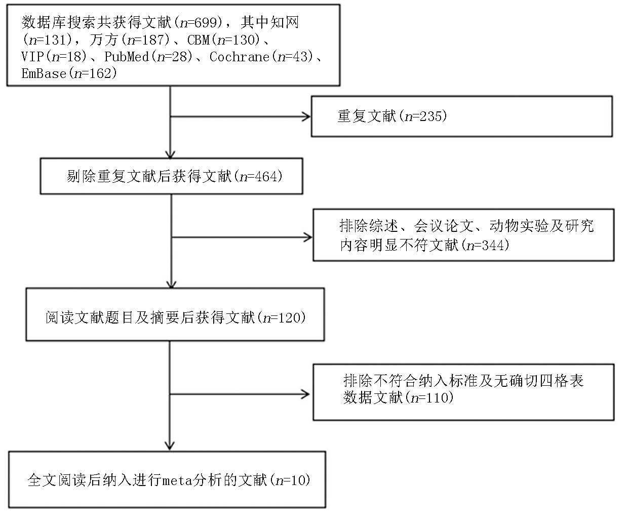
Fig.1 Flowchart for literature screen
| 第一作者 | 发表 年份 | 不同国家的 超声机器 | 研究对象 | 脑卒中诊断标准 | RR(95%CI) |
|---|---|---|---|---|---|
| 顾秀娟等[ | 2022 | Toshiba(日本) | 经超声证实的颈动脉斑块致狭窄(狭窄率≥50%)患者 | 近6个月内有无同侧同流域缺血性脑卒中症状 | 5.33(3.07, 9.24) |
| 李海欣等[ | 2021 | Toshiba(日本) | 颈动脉斑块患者 | 6个月内有无短暂性脑缺血发作或非致死性脑卒中 | 2.83(1.67, 4.78) |
| Jain等[ | 2020 | GE(美国) | 颈动脉斑块致狭窄(狭窄率≥60%)患者 | 有或没有缺血性脑血管症状 | 1.19(0.74, 1.91) |
| 韦玉亚等[ | 2022 | NA | 接受颈动脉内膜切除术治疗的患者 | 有无短暂性脑缺血或缺血性脑卒中症状 | 2.62(1.56, 4.38) |
| Zhao等[ | 2022 | Mindray(中国) | 急性脑梗死患者及颈动脉斑块患者 | CT或MRI | 5.42(3.31, 8.88) |
| Cui等[ | 2021 | Toshiba(日本) | 无症状颈动脉斑块狭窄患者 | CT或MRI | 3.27(1.23, 8.66) |
| Li等[ | 2019 | Siemens(德国) | 短暂性脑缺血发作(TIA)患者 | 随访获得 | 2.04(1.26, 3.29) |
| 孙飞一等[ | 2022 | SuperSonic Imagine(法国) | 短暂性脑缺血发作(TIA)患者 | 随访获得 | 2.59(1.48, 4.50) |
| 唐诗琪等[ | 2021 | HITACHI(日本) | 急性非心源性缺血性脑卒中患者 | CT或MRI | 4.15(2.78, 6.18) |
| Song等[ | 2021 | GE(美国) | 近期发生脑卒中且至少有一个颈动脉粥样硬化斑块的患者 | 随访 | 7.70(4.06, 14.59) |
Tab.1 Basic characteristics of the included literatures
| 第一作者 | 发表 年份 | 不同国家的 超声机器 | 研究对象 | 脑卒中诊断标准 | RR(95%CI) |
|---|---|---|---|---|---|
| 顾秀娟等[ | 2022 | Toshiba(日本) | 经超声证实的颈动脉斑块致狭窄(狭窄率≥50%)患者 | 近6个月内有无同侧同流域缺血性脑卒中症状 | 5.33(3.07, 9.24) |
| 李海欣等[ | 2021 | Toshiba(日本) | 颈动脉斑块患者 | 6个月内有无短暂性脑缺血发作或非致死性脑卒中 | 2.83(1.67, 4.78) |
| Jain等[ | 2020 | GE(美国) | 颈动脉斑块致狭窄(狭窄率≥60%)患者 | 有或没有缺血性脑血管症状 | 1.19(0.74, 1.91) |
| 韦玉亚等[ | 2022 | NA | 接受颈动脉内膜切除术治疗的患者 | 有无短暂性脑缺血或缺血性脑卒中症状 | 2.62(1.56, 4.38) |
| Zhao等[ | 2022 | Mindray(中国) | 急性脑梗死患者及颈动脉斑块患者 | CT或MRI | 5.42(3.31, 8.88) |
| Cui等[ | 2021 | Toshiba(日本) | 无症状颈动脉斑块狭窄患者 | CT或MRI | 3.27(1.23, 8.66) |
| Li等[ | 2019 | Siemens(德国) | 短暂性脑缺血发作(TIA)患者 | 随访获得 | 2.04(1.26, 3.29) |
| 孙飞一等[ | 2022 | SuperSonic Imagine(法国) | 短暂性脑缺血发作(TIA)患者 | 随访获得 | 2.59(1.48, 4.50) |
| 唐诗琪等[ | 2021 | HITACHI(日本) | 急性非心源性缺血性脑卒中患者 | CT或MRI | 4.15(2.78, 6.18) |
| Song等[ | 2021 | GE(美国) | 近期发生脑卒中且至少有一个颈动脉粥样硬化斑块的患者 | 随访 | 7.70(4.06, 14.59) |
| 纳入研究 | 发表年份 | 选择 | 可比性 | 结局 | 得分 |
|---|---|---|---|---|---|
| 顾秀娟等[ | 2022 | **** | ** | *** | 9 |
| 李海欣等[ | 2021 | **** | * | *** | 8 |
| Jain等[ | 2020 | *** | * | ** | 6 |
| 韦玉亚等[ | 2022 | *** | * | ** | 6 |
| Zhao等[ | 2022 | **** | * | *** | 8 |
| Cui等[ | 2021 | *** | ** | ** | 7 |
| Li等[ | 2019 | **** | * | *** | 8 |
| 孙飞一等[ | 2022 | *** | * | *** | 7 |
| 唐诗琪等[ | 2021 | *** | ** | *** | 8 |
| Song等[ | 2021 | **** | * | *** | 8 |
Tab.2 Results of the literature quality assessment
| 纳入研究 | 发表年份 | 选择 | 可比性 | 结局 | 得分 |
|---|---|---|---|---|---|
| 顾秀娟等[ | 2022 | **** | ** | *** | 9 |
| 李海欣等[ | 2021 | **** | * | *** | 8 |
| Jain等[ | 2020 | *** | * | ** | 6 |
| 韦玉亚等[ | 2022 | *** | * | ** | 6 |
| Zhao等[ | 2022 | **** | * | *** | 8 |
| Cui等[ | 2021 | *** | ** | ** | 7 |
| Li等[ | 2019 | **** | * | *** | 8 |
| 孙飞一等[ | 2022 | *** | * | *** | 7 |
| 唐诗琪等[ | 2021 | *** | ** | *** | 8 |
| Song等[ | 2021 | **** | * | *** | 8 |
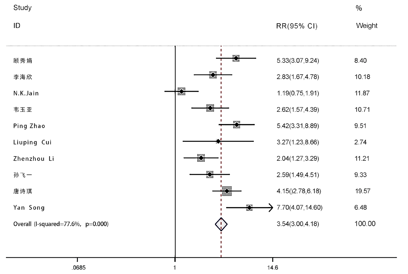
Fig.2 Correlation of CEUS assessment results with stroke occurrence
| 变量 | 纳入研究数 | 异质性检验 | RR( 95%CI) | P值 | |
|---|---|---|---|---|---|
| I2 值(%) | P值 | ||||
| 样本量 | 0.002 | ||||
| >100 | 5 | 22.2 | 0.273 | 4.08 (3.13, 5.31) | |
| <100 | 5 | 43.0 | 0.135 | 2.09 (1.51, 2.91) | |
| 诊断标准 | 0.044 | ||||
| 临床症状 | 4 | 82.6 | <0.001 | 2.62 (1.42, 4.84) | |
| CT或MRI | 3 | 0.0 | 0.571 | 4.46 (3.32, 6.00) | |
| 随访 | 3 | 0.0 | 0.730 | 2.34 (1.62, 3.24) | |
| 研究对象 | 0.806 | ||||
| 有脑卒中既往史患者 | 4 | 46.0 | 0.135 | 2.85 (1.97, 4.97) | |
| 无脑卒中既往史颈动脉斑块患者 | 1 | - | 0.000 | 3.26 (1.23, 8.60) | |
| 超声仪器生产厂家 | 0.082 | ||||
| Toshiba | 3 | 0.0 | 0.400 | 3.91 (3.00, 5.10) | |
| 其他 | 6 | 74.6 | 0.001 | 2.50 (1.63, 3.84) | |
Tab.3 Subgroup analysis
| 变量 | 纳入研究数 | 异质性检验 | RR( 95%CI) | P值 | |
|---|---|---|---|---|---|
| I2 值(%) | P值 | ||||
| 样本量 | 0.002 | ||||
| >100 | 5 | 22.2 | 0.273 | 4.08 (3.13, 5.31) | |
| <100 | 5 | 43.0 | 0.135 | 2.09 (1.51, 2.91) | |
| 诊断标准 | 0.044 | ||||
| 临床症状 | 4 | 82.6 | <0.001 | 2.62 (1.42, 4.84) | |
| CT或MRI | 3 | 0.0 | 0.571 | 4.46 (3.32, 6.00) | |
| 随访 | 3 | 0.0 | 0.730 | 2.34 (1.62, 3.24) | |
| 研究对象 | 0.806 | ||||
| 有脑卒中既往史患者 | 4 | 46.0 | 0.135 | 2.85 (1.97, 4.97) | |
| 无脑卒中既往史颈动脉斑块患者 | 1 | - | 0.000 | 3.26 (1.23, 8.60) | |
| 超声仪器生产厂家 | 0.082 | ||||
| Toshiba | 3 | 0.0 | 0.400 | 3.91 (3.00, 5.10) | |
| 其他 | 6 | 74.6 | 0.001 | 2.50 (1.63, 3.84) | |
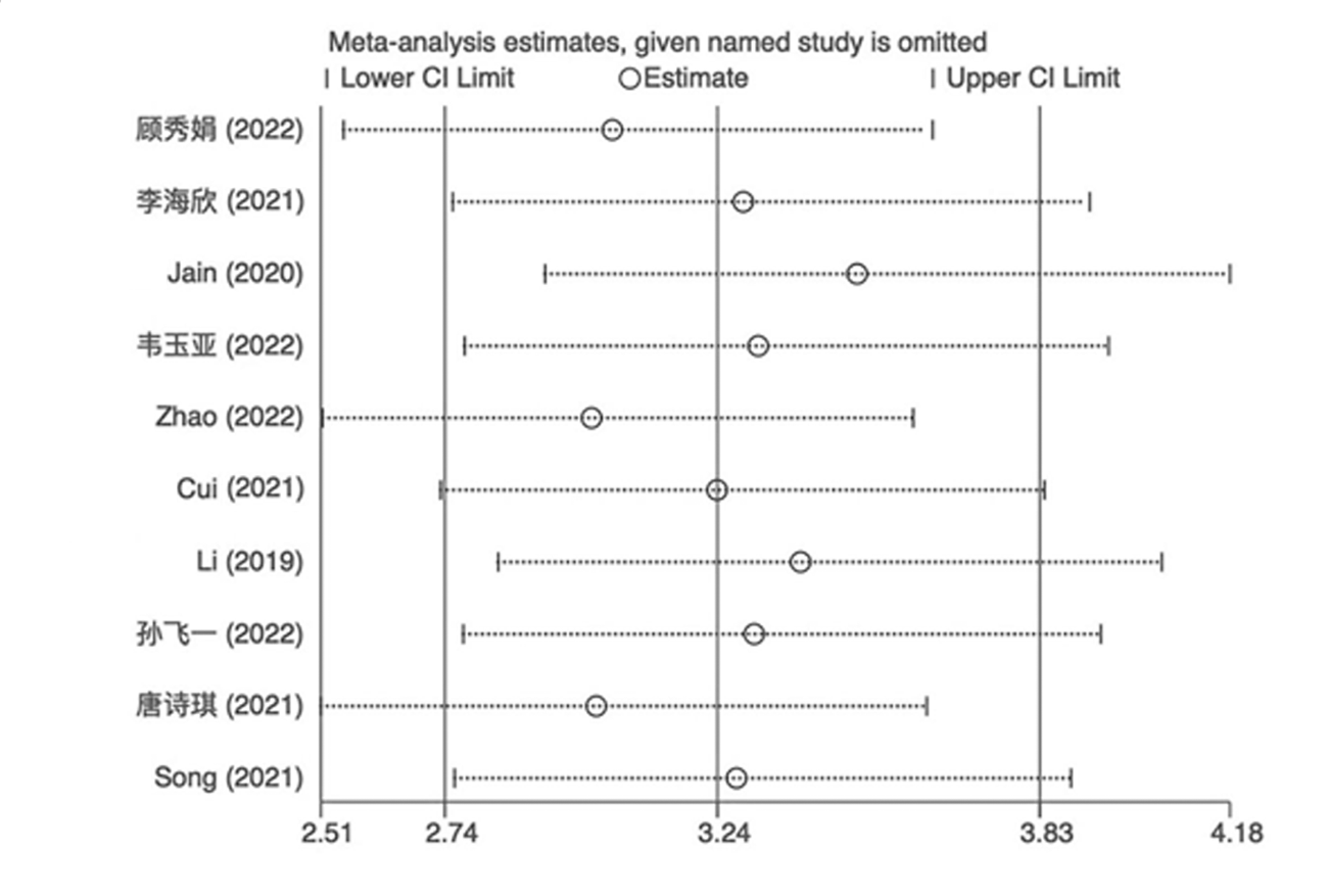
Fig.3 Sensitivity analysis
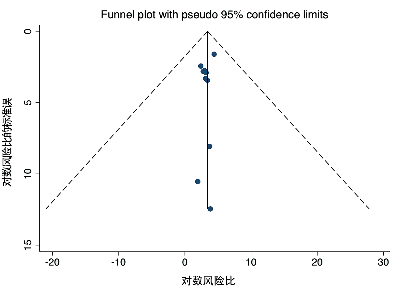
Fig.4 Funnel plots for publication bias analysis
| [1] |
Fleg JL, Stone GW, Fayad ZA, et al. Detection of high-risk atherosclerotic plaque: Report of the NHLBI Working Group on current status and future directions[J]. JACC Cardiovasc Imaging, 2012, 5(9): 941-955.
doi: 10.1016/j.jcmg.2012.07.007 pmid: 22974808 |
| [2] |
Ritman EL, Lerman A. The dynamic vasa vasorum[J]. Cardiovasc Res, 2007, 75(4): 649-658.
doi: 10.1016/j.cardiores.2007.06.020 pmid: 17631284 |
| [3] | Michel JB, Martin-Ventura JL, Nicoletti A, et al. Pathology of human plaque vulnerability: Mechanisms and consequences of intraplaque haemorrhages[J]. Atherosclerosis, 2014, 234(2): 311-319. |
| [4] | 顾秀娟, 张巍, 郑帅, 等. 颈动脉斑块常规超声及超声造影预测颈动脉狭窄患者缺血性脑卒中[J]. 中国介入影像与治疗学, 2022, 19(9): 570-574. |
| [5] | 李海欣, 阚艳敏, 宁彬, 等. 颈动脉斑块超声造影增强特征与其病理特征及易损性的相关性[J]. 中国医学影像技术, 2021, 37(6): 857-861. |
| [6] | Jain NK, Singh G, Muralidharan CG, et al. Assessment of plaque vulnerability in carotid atherosclerotic plaques using contrast-enhanced ultrasound[J]. Med J Armed Forces India., 2020, 78 (4): 422-429. |
| [7] | 韦玉亚, 朱婷, 方静, 等. 多模态超声检查技术诊断颈动脉斑块稳定性的价值[J]. 现代医用影像学, 2022, 31(6): 1175-1178,1188. |
| [8] | Zhao P, Xu E, Yuan R, et al. The predictive value of contrast-enhanced ultrasound combined with serum miR-124 level in acute cerebral infarction and their correlation with the contrast enhancement of carotid atherosclerotic plaque[J]. Neuropsychiatr Dis Treat, 2022, 11(18): 1397-1403. |
| [9] | Cui L, Xing Y, Zhou Y, et al. Carotid intraplaque neovascularisation as a predictive factor for future vascular events in patients with mild and moderate carotid stenosis: An observational prospective study[J]. Ther Adv Neurol Disord, 2021, 19(14): 17562864211023992. |
| [10] | Li Z, Xu X, Ren L, et al. Prospective study about the relationship between CEUS of carotid intraplaque neovascularization and ischemic stroke in TIA patients[J]. Front Pharmacol, 2019, 21(10): 672. |
| [11] | 孙飞一, 潘晓芳, 贾晓东, 等. 多模态超声检查结合临床预测颈动脉粥样硬化患者缺血性脑卒中或短暂性脑缺血复发[J]. 中国医学影像技术, 2022, 38(1): 53-58. |
| [12] | 唐诗琪, 李涛. 超声造影评估颈动脉斑块内新生血管与脑梗死再发的相关性[J]. 中国社区医师, 2021, 37(22):96-97. |
| [13] |
Song Y, Dang Y, Wang J, et al. Carotid intraplaque neovascularization predicts ischemic stroke recurrence in patients with carotid atherosclerosis[J]. Gerontology, 2021, 67(2): 144-151.
doi: 10.1159/000511360 pmid: 33582668 |
| [14] |
Fleiner M, Kununer M, Mirlacher M, et al. Arterial neovascularization and inflammation in vulnerable patients: Early and late signs of symptomatic atherosclerosis[J]. Circulation, 2004, 110(18): 2843-2850.
pmid: 15505090 |
| [15] | 刘学钧, 刘奕蓉, 周建平. 动脉粥样硬化斑块与老年脑卒中的相关性[J]. 中国老年学杂志, 2010, 30(3):395-396. |
| [16] | 于晶, 侯苏芸, 张艳. 超声造影对颈动脉斑块稳定性的价值探讨[J]. 中国实用神经疾病杂志, 2018, 21(7): 757-760. |
| [17] | 张学谦, 田晖, 贾丽景, 等. 常规超声与超声造影诊断颈动脉粥样硬化斑块的对比研究[J]. 临床荟萃, 2016, 31(11): 1233-1236. |
| [18] | Baun J. Contrast-enhanced ultrasound: Identification of neovascular-ization permits characterization of vulnerable carotid plaques[J]. JDMS, 2020, 36: 471-477. |
| [19] | 中华医学会神经病学分会, 中华医学会神经病学分会脑血管病学组. 中国急性缺血性卒中诊治指南2023[J]. 中华神经科杂志, 2024, 57(6): 523-559. |
| [20] | 罗彤, 高阳, 何金龙, 等. 磁共振成像技术和病因重叠在缺血性脑卒中复发风险预测中的研究进展[J]. 磁共振成像, 2023, 14(10): 152-156,166. |
| Viewed | ||||||
|
Full text |
|
|||||
|
Abstract |
|
|||||