Clinical Focus ›› 2026, Vol. 41 ›› Issue (9): 794-806.doi: 10.3969/j.issn.1004-583X.2026.09.003
Previous Articles Next Articles
Differences in the gut microbiota among patients with benign and malignant pulmonary nodules and healthy individuals
Xie Shiliang1,2a, Wang Xinyu3, Zhao Long4, Sun Ruixin2b( )
- 1
Health Science Center ,Ningbo University Ningbo 315211, China
2Department of Cardiothoracic Surgery; b.Department of Clinical Laboratory ,Shanghai Tongji Hospital Shanghai 200065, China
3Shanghai University of Medicine & Health Sciences Shanghai 201318, China
4Department of Cardiac and Great Vascular Surgery ,the Second Affiliated Hospital of Zhejiang University School of Medicine Hangzhou 310009, China
-
Received:2026-05-20Online:2026-09-20Published:2026-09-17 -
Contact:Sun Ruixin E-mail:sunruixin@tongji.edu.cn

CLC Number:
Cite this article
Xie Shiliang, Wang Xinyu, Zhao Long, Sun Ruixin. Differences in the gut microbiota among patients with benign and malignant pulmonary nodules and healthy individuals[J]. Clinical Focus, 2026, 41(9): 794-806.
share this article
Add to citation manager EndNote|Ris|BibTeX
URL: https://www.lchc.cn/EN/10.3969/j.issn.1004-583X.2026.09.003
| 项目 | 恶性肺结节组 (n=23) | 良性肺结节组 (n=8) | 健康人群组 (n=14) | χ2/H值 | P值 |
|---|---|---|---|---|---|
| 年龄(岁) | 68.0(58.0, 74.0)* | 59.5(55.0, 65.5) | 50.5(42.0, 61.8) | 9.615 | 0.008 |
| 男性[例(%)] | 13(56.5) | 4(50.0) | 7(50.0) | 0.192 | 0.908 |
| 体质量指数(kg/m2) | 24.02(21.18, 25.72) | 24.87(21.05, 26.95) | 23.84(23.37, 26.51) | 1.004 | 0.606 |
| 吸烟史[例(%)] | 5(21.7) | 2(25.0) | 4(28.6) | 0.222 | 0.895 |
| 饮酒史[例(%)] | 2(8.7) | 1(12.5) | 2(14.3) | 0.294 | 0.863 |
| 合并基础疾病[例(%)] | 9(39.1) | 1(12.5) | 3(21.4) | 2.600 | 0.273 |
| 结节最大径[例(%)] | |||||
| ≤1.0 cm | 11(47.8) | - | - | - | - |
| >1.0 cm | 12(52.2) | - | - | - | - |
| 分化程度[例(%)] | |||||
| 高分化 | 12(52.2) | - | - | - | - |
| 中分化 | 7(30.4) | - | - | - | - |
| 低分化 | 4(17.4) | - | - | - | - |
| 有无淋巴结转移[例(%)] | |||||
| 有 | 0 | - | - | - | - |
| 无 | 23(100.0) | - | - | - | - |
Tab.1 Baseline characteristics of the three groups and clinical stratification of the malignant pulmonary nodule group
| 项目 | 恶性肺结节组 (n=23) | 良性肺结节组 (n=8) | 健康人群组 (n=14) | χ2/H值 | P值 |
|---|---|---|---|---|---|
| 年龄(岁) | 68.0(58.0, 74.0)* | 59.5(55.0, 65.5) | 50.5(42.0, 61.8) | 9.615 | 0.008 |
| 男性[例(%)] | 13(56.5) | 4(50.0) | 7(50.0) | 0.192 | 0.908 |
| 体质量指数(kg/m2) | 24.02(21.18, 25.72) | 24.87(21.05, 26.95) | 23.84(23.37, 26.51) | 1.004 | 0.606 |
| 吸烟史[例(%)] | 5(21.7) | 2(25.0) | 4(28.6) | 0.222 | 0.895 |
| 饮酒史[例(%)] | 2(8.7) | 1(12.5) | 2(14.3) | 0.294 | 0.863 |
| 合并基础疾病[例(%)] | 9(39.1) | 1(12.5) | 3(21.4) | 2.600 | 0.273 |
| 结节最大径[例(%)] | |||||
| ≤1.0 cm | 11(47.8) | - | - | - | - |
| >1.0 cm | 12(52.2) | - | - | - | - |
| 分化程度[例(%)] | |||||
| 高分化 | 12(52.2) | - | - | - | - |
| 中分化 | 7(30.4) | - | - | - | - |
| 低分化 | 4(17.4) | - | - | - | - |
| 有无淋巴结转移[例(%)] | |||||
| 有 | 0 | - | - | - | - |
| 无 | 23(100.0) | - | - | - | - |
| 病理分型 | 例数 | CEA(μg/L) | NSE(μg/L) | CYFRA21-1(μg/L) | SCCA(μg/L) |
|---|---|---|---|---|---|
| 腺癌 | 18 | 2.885(2.145, 4.660) | 12.000(11.200, 16.650) | 2.280(1.768, 2.700) | 1.105(0.843, 1.458) |
| 非腺癌 | 5 | 2.410(0.970, 2.740) | 11.000(9.130, 13.100) | 3.370(2.880, 4.170) | 2.110(0.820, 3.070) |
| Z值 | -1.342 | -0.895 | -1.715 | -1.006 | |
| P值 | 0.180 | 0.371 | 0.086 | 0.314 |
Tab.2 Comparison of serum biomarker levels between patients with adenocarcinoma and non-adenocarcinoma
| 病理分型 | 例数 | CEA(μg/L) | NSE(μg/L) | CYFRA21-1(μg/L) | SCCA(μg/L) |
|---|---|---|---|---|---|
| 腺癌 | 18 | 2.885(2.145, 4.660) | 12.000(11.200, 16.650) | 2.280(1.768, 2.700) | 1.105(0.843, 1.458) |
| 非腺癌 | 5 | 2.410(0.970, 2.740) | 11.000(9.130, 13.100) | 3.370(2.880, 4.170) | 2.110(0.820, 3.070) |
| Z值 | -1.342 | -0.895 | -1.715 | -1.006 | |
| P值 | 0.180 | 0.371 | 0.086 | 0.314 |
| 病理分期 | 例数 | CEA(μg/L) | NSE(μg/L) | CYFRA21-1(μg/L) | SCCA(μg/L) |
|---|---|---|---|---|---|
| Ⅰ期 | 19 | 2.890(2.160, 4.620) | 11.600(10.800, 16.100) | 2.310(1.835, 2.765) | 1.080(0.805, 1.535) |
| Ⅱ期 | 4 | 1.745(1.052, 2.493) | 12.050(10.430, 16.950) | 3.770(2.765, 4.452) | 1.620(1.052, 2.430) |
| Z值 | -1.886 | -1.785 | -1.460 | -0.284 | |
| P值 | 0.062 | 0.074 | 0.144 | 0.776 |
Tab.3 Comparison of serum biomarker levels among patients with lung cancer at different pathological stages
| 病理分期 | 例数 | CEA(μg/L) | NSE(μg/L) | CYFRA21-1(μg/L) | SCCA(μg/L) |
|---|---|---|---|---|---|
| Ⅰ期 | 19 | 2.890(2.160, 4.620) | 11.600(10.800, 16.100) | 2.310(1.835, 2.765) | 1.080(0.805, 1.535) |
| Ⅱ期 | 4 | 1.745(1.052, 2.493) | 12.050(10.430, 16.950) | 3.770(2.765, 4.452) | 1.620(1.052, 2.430) |
| Z值 | -1.886 | -1.785 | -1.460 | -0.284 | |
| P值 | 0.062 | 0.074 | 0.144 | 0.776 |
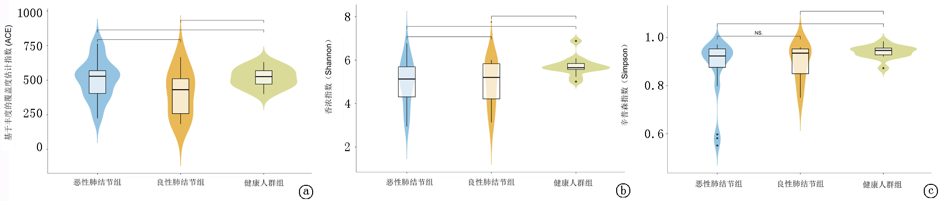
Fig.1 Alpha diversity indices of the three groups a. ACE index; b. Shannon index; c. Simpson index.Note: The x-axis represents the group, and the y-axis represents the corresponding diversity index value. The black lines above the plots indicate a statistically significant difference between the two groups (P<0.05), whereas black lines labeled NS indicate no statistically significant difference between the two groups (P>0.05)
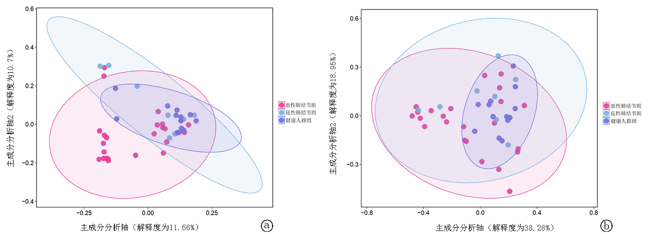
Fig.2 PCoA analysis of the three groups a. PCoA based on unweighted UniFrac distance; b. PCoA based on weighted UniFrac distance.Note: The x-axis represents the first principal component, and the y-axis represents the second principal component. The percentages indicate the contribution of the corresponding principal components to sample variation. Each dot represents a sample; different colors represent different groups; and the ellipse indicates the 95% confidence region of each group

Fig.3 LEfSe analysis of the malignant pulmonary nodule group and the healthy control group a. LDA score histogram (with |LDA score| >3 as the threshold); the x-axis indicates the LDA scores of each taxon, and the y-axis indicates the taxa with statistically significant intergroup differences; longer bars indicate more significant differences; b. Evolutionary cladogram, with the inner-to-outer rings representing taxa from phylum to genus; the diameter of each circle at different taxonomic levels is proportional to its relative abundance
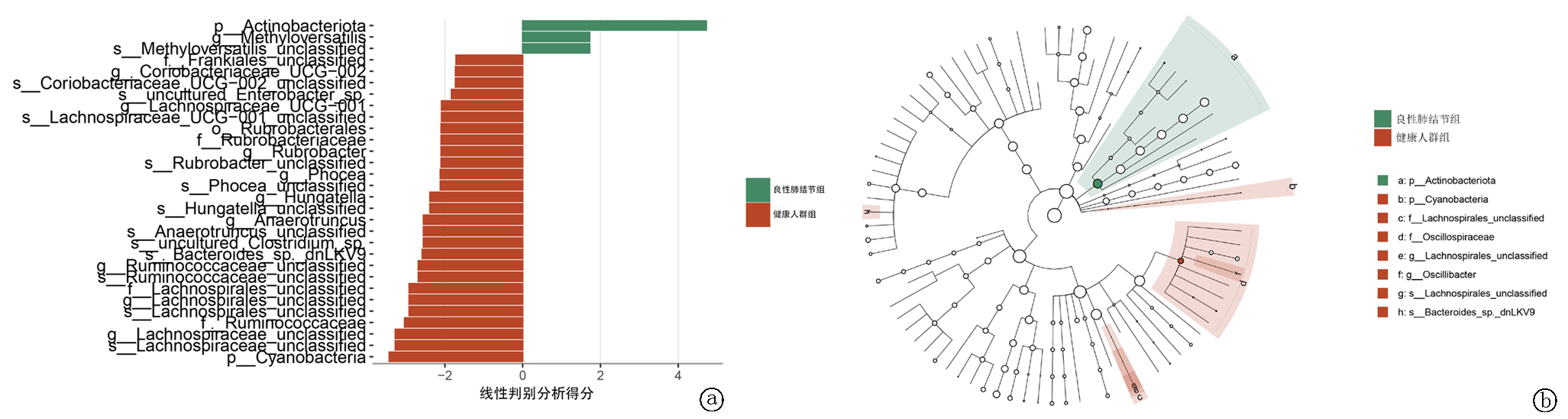
Fig.4 LEfSe analysis of the benign pulmonary nodule group and the healthy control group: a. LDA score histogram; b. Evolutionary cladogram
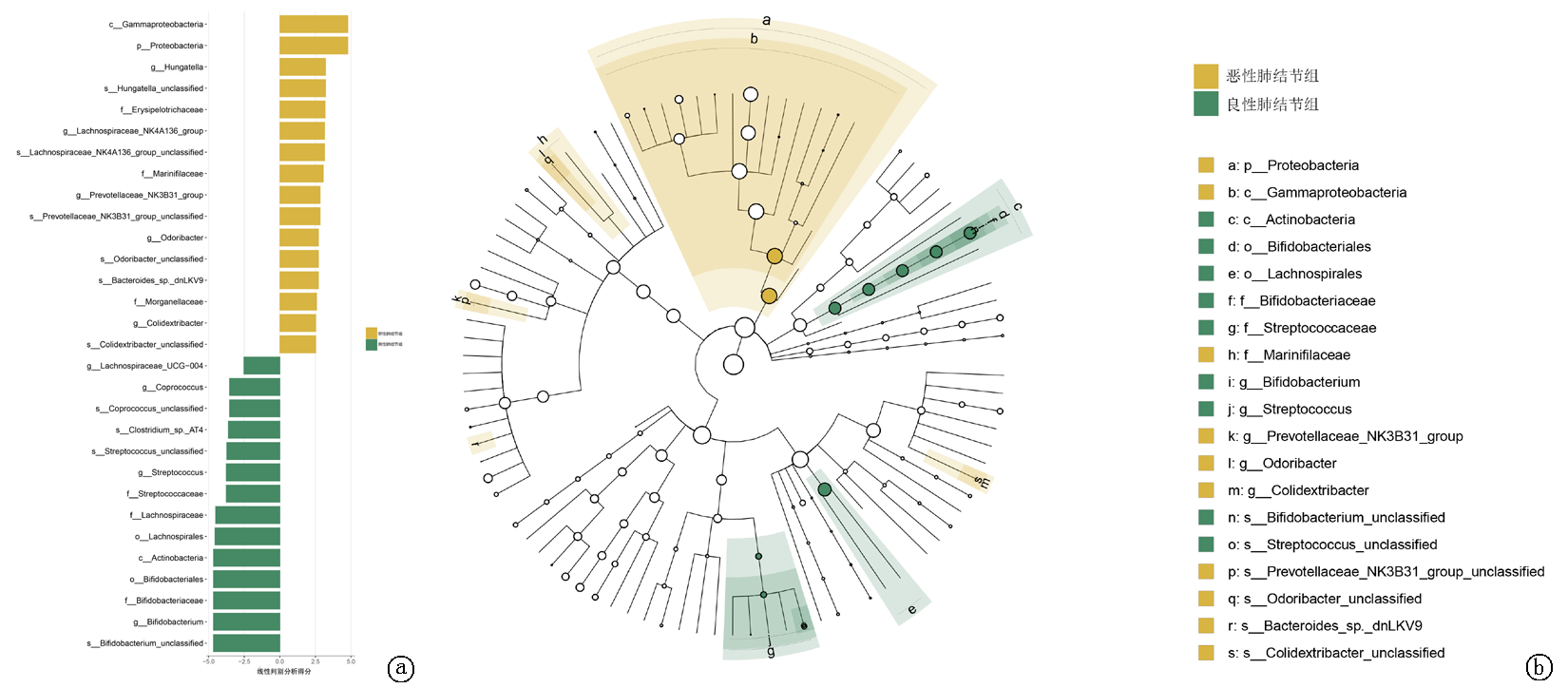
Fig.5 LEfSe analysis of the malignant pulmonary nodule group and the benign pulmonary nodule group a. LDA score histogram; b. Evolutionary cladogram
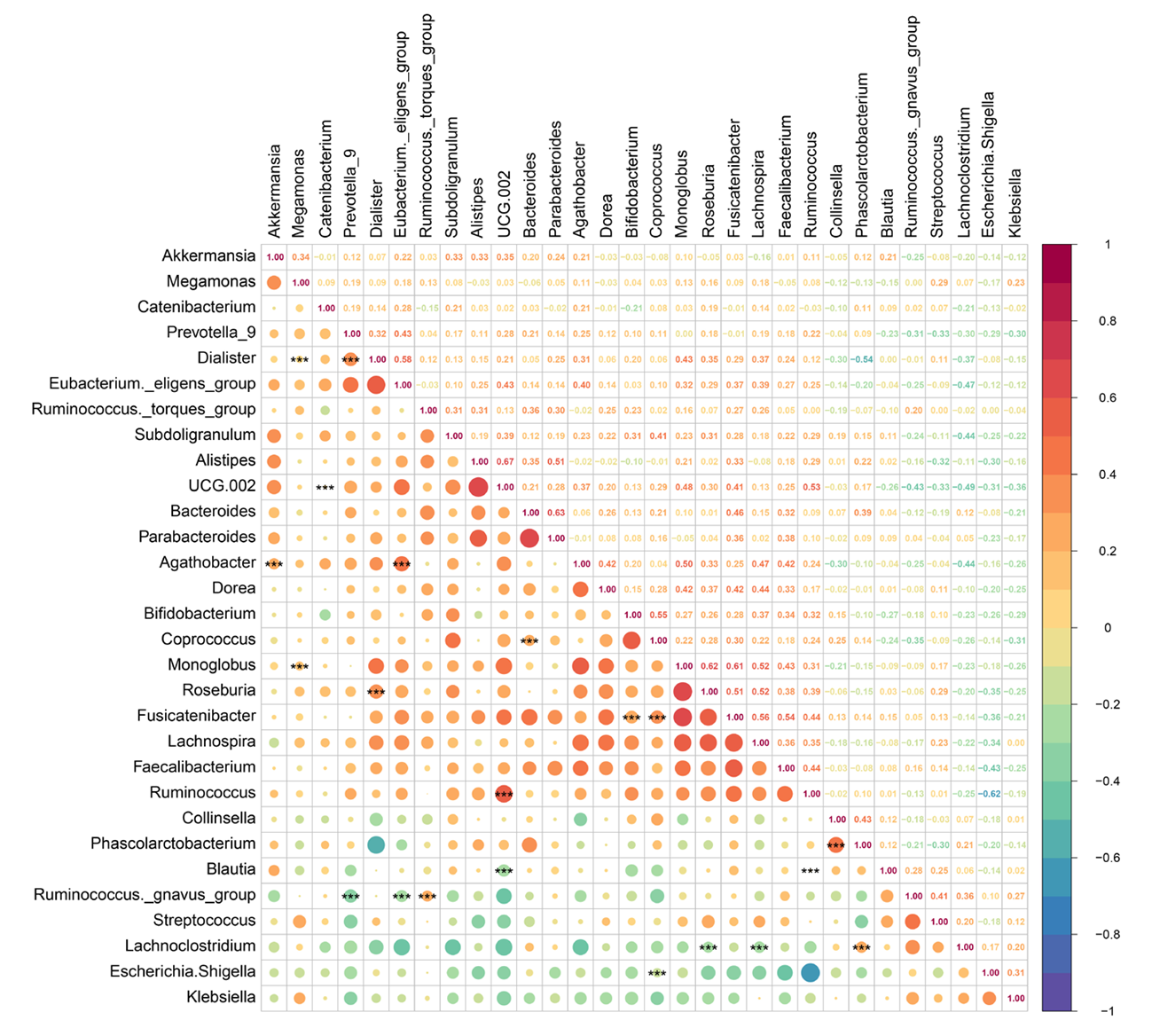
Fig.6 Correlation heatmap at the genus level Note: The x-axis and y-axis list the genus names of various bacteria, and the color bar on the right indicates values ranging from -1 to 1, representing the correlation between two genera. Values closer to -1 indicate a stronger negative correlation, whereas values closer to 1 indicate a stronger positive correlation. ***: P<0.01
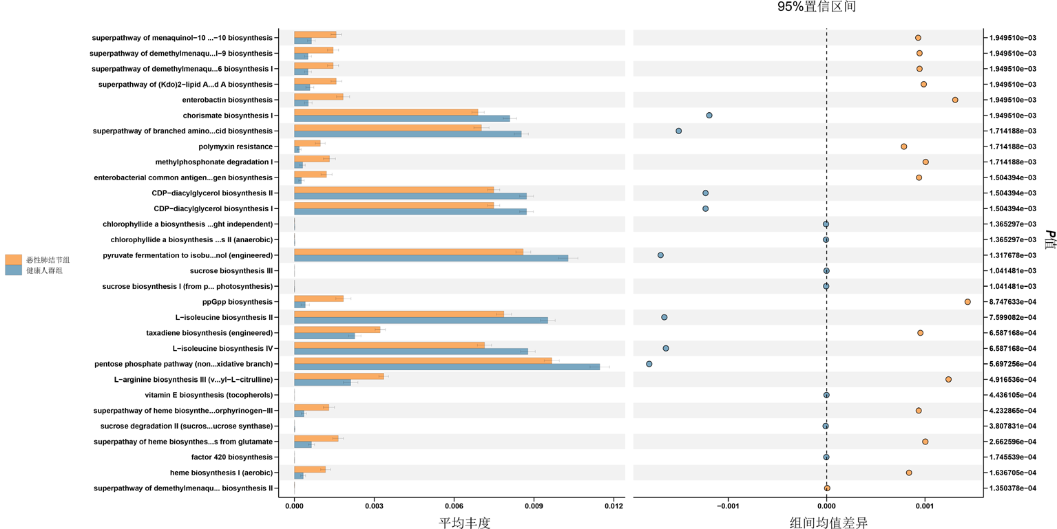
Fig.7 Differential analysis of KEGG pathways between the malignant pulmonary nodule group and the healthy control group Note: The x-axis represents the relative abundance percentage of metabolic pathways, and the y-axis represents the pathway names. The figure shows differences under the 95% confidence interval; the farther from the center line, the greater the difference
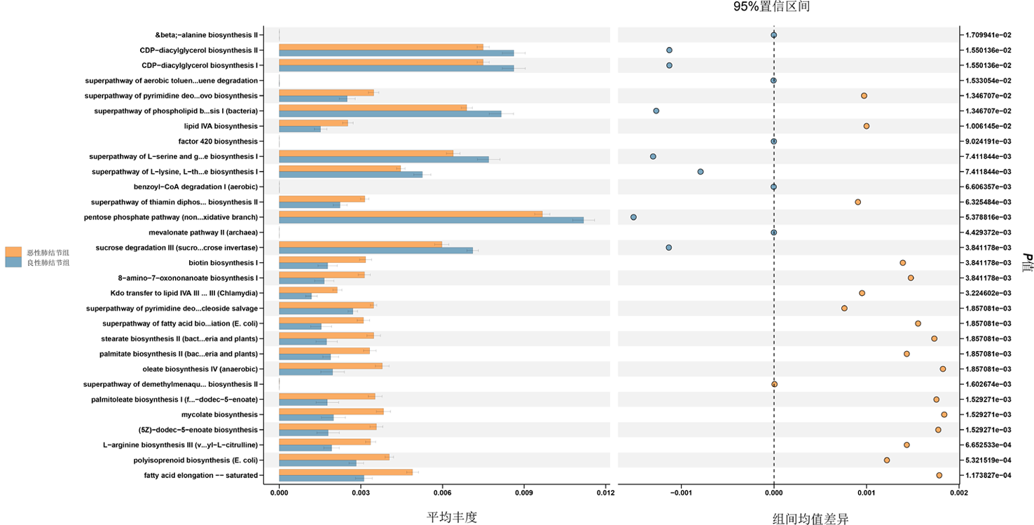
Fig.8 Differential KEGG pathways between the malignant pulmonary nodule group and the benign pulmonary nodule group
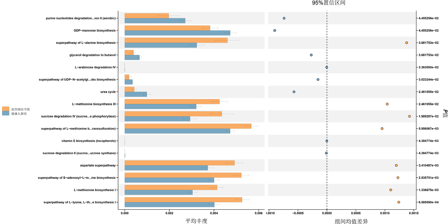
Fig.9 Differential KEGG pathways between the benign pulmonary nodule group and the healthy control group
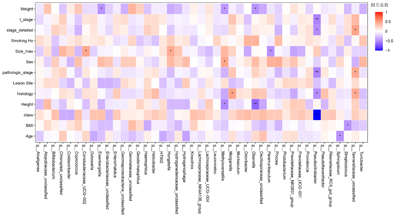
Fig.10 Clinical information heatmap Note: The x-axis represents bacterial genera, and the y-axis represents related clinical information, including core clinical variables such as body measurements, disease stage, clinical behavior, tumor morphology, and age. A correlation coefficient closer to 1 (higher red saturation) indicates a stronger positive correlation, whereas a correlation coefficient closer to -1 (higher blue saturation) indicates a stronger negative correlation. *:P<0.05, **: P<0.01
| [1] | Bray F, Laversanne M, Sung H, et al. Global cancer statistics 2022: GLOBOCAN estimates of incidence and mortality worldwide for 36 cancers in 185 countries[J]. CA Cancer J Clin, 2024, 74(3): 229-263. doi: 10.3322/caac.21834. |
| [2] | George JE, George PS, Krishna JKM, et al. Global trends in lung cancer incidence and mortality by age, gender and morphology and forecast: A bootstrap-based analysis[J]. Lung Cancer, 2025, 205: 108626. doi: 10.1016/j.lungcan.2024.107954. |
| [3] | Oser MG, Niederst MJ, Sequist LV, et al. Transformation from non-small-cell lung cancer to small-cell lung cancer: Molecular drivers and cells of origin[J]. Lancet Oncol, 2015, 16(4): e165-e172. doi: 10.1016/S1470-2045(14)71180-5. |
| [4] |
Thai AA, Solomon BJ, Sequist LV, et al. Lung cancer[J]. Lancet, 2021, 398(10299): 535-554. doi: 10.1016/S0140-6736(21)00124-4.
pmid: 34273294 |
| [5] | Li Y, Yan B, He S. Advances and challenges in the treatment of lung cancer[J]. Biomed Pharmacother, 2023, 169: 115891. doi: 10.1016/j.biopha.2023.115891. |
| [6] | Bi JH, Tuo JY, Xiao YX, et al. Observed and relative survival trends of lung cancer: A systematic review of population-based cancer registration data[J]. Thorac Cancer, 2024, 15(2): 142-151. doi: 10.1111/1759-7714.15163. |
| [7] |
Zhang J, Mj IJ, Oberoi J, et al. Time to diagnosis and treatment of lung cancer: A systematic overview of risk factors, interventions and impact on patient outcomes[J]. Lung Cancer, 2022, 166: 27-39. doi: 10.1016/j.lungcan.2022.01.014.
pmid: 35152171 |
| [8] | Puri N, Sahane P, Phatale V, et al. Nano-chameleons: A review on cluster of differentiation-driven immune cell-engineered nanoarchitectonics for non-small cell lung cancer[J]. Int J Biol Macromol, 2025, 310(Pt 4): 143440. doi: 10.1016/j.ijbiomac.2024.131120. |
| [9] | Duan YF, Dai JH, Lu YQ, et al. Disentangling the molecular mystery of tumour-microbiota interactions: Microbial metabolites[J]. Clin Transl Med, 2024, 14(11): e70093. doi: 10.1002/ctm2.70093. |
| [10] | Zhu B, Wang X, Li L. Human gut microbiome: the second genome of human body[J]. Protein Cell, 2010, 1(8): 718-725. doi: 10.1007/s13238-010-0093-z. |
| [11] | Khalil M, Di Ciaula A, Mahdi L, et al. Unraveling the role of the human gut microbiome in health and diseases[J]. Microorganisms, 2024, 12(11): 2154. doi: 10.3390/microorganisms12112154. |
| [12] | Islam MM, Mahbub NU, Hong ST, et al. Gut bacteria: An etiological agent in human pathological conditions[J]. Front Cell Infect Microbiol, 2024, 14: 1291148. doi: 10.3389/fcimb.2024.1291148. |
| [13] | Zhang T, Guo F, Zhang C, et al. Decoding the role of gut mycobiota in immune regulation and disease[J]. Virulence, 2025, 16(1): 2541704. doi: 10.1080/21505594.2024.2341704. |
| [14] | Lushchak VI. Symphony of digestion: coordinated host-microbiome enzymatic interplay in gut ecosystem[J]. Biomolecules, 2025, 15(8): 942. doi: 10.3390/biom15080942. |
| [15] | Cai J, Zhang W, Zhu S, et al. Gut and intratumoral microbiota: Key to lung cancer development and immunotherapy[J]. Int Immunopharmacol, 2025, 156: 114677. doi: 10.1016/j.intimp.2024.112577. |
| [16] | Smolarz B, Łukasiewicz H, Samulak D, et al. Lung cancer-epidemiology, pathogenesis, treatment and molecular aspect (review of literature)[J]. Int J Mol Sci, 2025, 26(5): 2754. doi: 10.3390/ijms26052754. |
| [17] | Ji Y, Zhang Y, Liu S, et al. The epidemiological landscape of lung cancer: Current status, temporal trend and future projections based on the latest estimates from GLOBOCAN 2022[J]. J Natl Cancer Cent, 2025, 5(3): 278-286. doi: 10.1016/j.jncc.2025.03.002. |
| [18] | Zhang R, Zhang X, Lau HCH, et al. Gut microbiota in cancer initiation, development and therapy[J]. Sci China Life Sci, 2025, 68(5): 1283-1308. doi: 10.1007/s11427-024-2451-4. |
| [19] | Min S, Zhang Y, Zhang H, et al. Gut microbiota drives cancer evolution and therapy resistance[J]. Mol Cancer, 2026, 25(1): 39. doi: 10.1186/s12943-026-01951-w. |
| [20] | Li X, Jia Y, Li Y, et al. Crosstalk between metabolic reprogramming and microbiota: Implications for cancer progression and novel therapeutic opportunities[J]. Front Immunol, 2025, 16: 1582166. doi: 10.3389/fimmu.2025.1582166. |
| [21] | Kim S, Seo SU, Kweon MN. Gut microbiota-derived metabolites tune host homeostasis fate[J]. Semin Immunopathol, 2024, 46(1-2): 2. doi: 10.1007/s00281-023-01002-3. |
| [22] | Chen Y, Xiao L, Zhou M, et al. The microbiota: A crucial mediator in gut homeostasis and colonization resistance[J]. Front Microbiol, 2024, 15: 1417864. doi: 10.3389/fmicb.2024.1417864. |
| [23] | Wang Q, Meng Q, Chen Y, et al. Interaction between gut microbiota and immunity in health and intestinal disease[J]. Front Immunol, 2025, 16: 1673852. doi: 10.3389/fimmu.2025.1673852. |
| [24] | Yu X, Yu X, Wang Y, et al. Respiratory diseases and the gut microbiota: An updated review[J]. Front Cell Infect Microbiol, 2025, 15: 1629005. doi: 10.3389/fcimb.2025.1629005. |
| [25] | Dora D, Szöcs E, Soós Á, et al. From bench to bedside: An interdisciplinary journey through the gut-lung axis with insights into lung cancer and immunotherapy[J]. Front Immunol, 2024, 15: 1434804. doi: 10.3389/fimmu.2024.1434804. |
| [26] | Hrncir T. Gut microbiota dysbiosis: Triggers, consequences, diagnostic and therapeutic options[J]. Microorganisms, 2022, 10(3): 578. doi: 10.3390/microorganisms10030578. |
| [27] |
Yang Y, Shen J, Wei S, et al. Characteristics of gut and lung microbiota in patients with lung masses and their relationship with clinical features[J]. BMC Microbiol, 2025, 25(1): 541. doi: 10.1186/s12866-025-03741-z.
pmid: 40849619 |
| [28] | Sha G, Wu Z, Wang B, et al. Intratumorally specific microbial-derived lipopolysaccharide contributes to non-small cell lung cancer progression[J]. Virulence, 2025, 16(1): 2548626. doi: 10.1080/21505594.2024.2348626. |
| [29] | Xu T, Wu X, Liu J, et al. The regulatory roles of dietary fibers on host health via gut microbiota-derived short chain fatty acids[J]. Curr Opin Pharmacol, 2022, 62: 36-42. doi: 10.1016/j.coph.2021.11.001. |
| [30] |
Rahal Z, Liu Y, Peng F, et al. Inflammation mediated by gut microbiome alterations promotes lung cancer development and an immunosuppressed tumor microenvironment[J]. Cancer Immunol Res, 2024, 12(12): 1736-1752. doi: 10.1158/2326-6066.CIR-24-0312.
pmid: 39269772 |
| [31] | Matoba R, Iijima H, Sakamoto Y, et al. Metabolic and functional pathways of gut microbiota in patients with gastric cancer[J]. Sci Rep, 2026, 16(1): 1432. doi: 10.1038/s41598-026-05188-x. |
| [1] | Chen Huimin, Ding Ruijuan, Liu He. Risk factors for comorbid anxiety and depression and for a single emotional disorder in patients with lung cancer receiving chemoradiotherapy [J]. Clinical Focus, 2026, 41(8): 736-741. |
| [2] | Tang Siying, Jia Junping, Tang Yuting, Zheng Yuqin, Kong Yue. Dietary inflammatory index and lung cancer risk: A meta-analysis [J]. Clinical Focus, 2026, 41(3): 197-204. |
| [3] | Qi Yumin, Wang Youjun. Correlation between serum bilirubin level and the imbalance of the normal gut microbiota in neonates with cholestatic jaundice [J]. Clinical Focus, 2022, 37(12): 1117-1121. |
| [4] | Huang Ping, Liu Hanyun, Li Yuhong. Risk factors of lung cancer with pulmonary embolism:a meta-analysis [J]. Clinical Focus, 2021, 36(12): 1061-1066. |
| [5] | Li Shuangping, Mao Yimin, He Xuegai. Diagnostic value of modified Wells score in lung cancer complicated with venous thromboembolism [J]. Clinical Focus, 2020, 35(5): 421-424. |
| [6] | Qiu Yi, Chen Lu, Qu Meidi, Zhang Min, Gu Hongliu, Sha Qi. Effect of nutritional status on nausea and vomiting caused by chemotherapy in patients with lung cancer [J]. Clinical Focus, 2019, 34(6): 549-552. |
| [7] | Yuan Boyun, Liu Xin, Yuan Yadong. Part of advances in respiratory disease during 2018 [J]. Clinical Focus, 2019, 34(1): 49-55. |
| [8] | Chen Benchao, Li Heng, Xiang Xudong, Li Gaofeng, Fu Feng, Guo Qi,Fan Shengliang,Wang Deguang. Efficacy and safety of icotinib combined with radiotherapy in the treatment of lung cancer with brain metastasis: a metaanalysis [J]. Clinical Focus, 2018, 33(8): 705-710. |
| [9] | Yuan Boyun, Gong Xiaowei, Yuan Yadong. Major advances in respiratory diseases during 2017 [J]. Clinical Focus, 2018, 33(1): 16-22. |
| [10] | Cai Jingjing1,2, Miao Jianlong1, Zhou Jinhua1, Zhang Yingying1,2, Liu Ruijuan1. Clinical features of chronic obstructive pulmonary disease with lung cancer [J]. Clinical Focus, 2017, 32(12): 1065-. |
| [11] | Chi Yan. Gut microbiota and inflammatory bowel disease [J]. Clinical Focus, 2016, 31(8): 857-860. |
| [12] | Liu Xinyan,Wei Hongyan, Dai Xianli, Liu Ying. Clinical efficacy of chemotherapy combined with location intervened by bronchoscope in central lung cancer [J]. Clinical Focus, 2016, 31(4): 389-391. |
| [13] | Yuan Yadong, Chen Ye,Wang Li,Shen Lixiao. Clinical advances in pulmonary during 2015 [J]. Clinical Focus, 2016, 31(2): 143-150. |
| [14] | Jin Faguang. Early diagnosis of lung cancer with new bronchoscopy techniques [J]. Clinical Focus, 2016, 31(11): 1161-1166. |
| [15] | Li Long;Tang Fabing;Yu Qin. Expression of EGFR and RAS/MAPK signal pathway-related proteins and their significance in lung adenocarcinoma [J]. Clinical Focus, 2015, 30(3): 290-292296. |
| Viewed | ||||||
|
Full text |
|
|||||
|
Abstract |
|
|||||