Clinical Focus ›› 2026, Vol. 41 ›› Issue (7): 619-625.doi: 10.3969/j.issn.1004-583X.2026.07.007
Previous Articles Next Articles
Clinical characteristics of dural arteriovenous fistula presenting primarily as epilepsy: Analysis of 4 cases
Dong Siyu1, Gao Tianwen2, Sun Mei2, Jin Shanquan2( )
)
- 1
Department of Neurology ,the First Affiliated Hospital of Wannan Medical University (Yijishan Hospital)
2Department of Neurology ,the First Affiliated Hospital of Nanjing Medical University
-
Received:2026-04-02Online:2026-07-20Published:2026-07-17 -
Contact:Jin Shanquan,Email:shanquanjin@qq.com

CLC Number:
Cite this article
Dong Siyu, Gao Tianwen, Sun Mei, Jin Shanquan. Clinical characteristics of dural arteriovenous fistula presenting primarily as epilepsy: Analysis of 4 cases[J]. Clinical Focus, 2026, 41(7): 619-625.
share this article
Add to citation manager EndNote|Ris|BibTeX
URL: https://www.lchc.cn/EN/10.3969/j.issn.1004-583X.2026.07.007
| 序号 | 性别 | 年龄 | 症状 | 既往史 | 头颅MRI | 脑血管造影 | 治疗 | 预后 |
|---|---|---|---|---|---|---|---|---|
| 1 | 男 | 57岁 | 视力下降,癫痫,帕金森 | 糖尿病、高血压病 | 右侧颞枕叶异常信号,血管源性水肿 | 右侧乙状窦动静脉瘘,右侧乙状窦重度狭窄,左侧乙状窦狭窄 | 经动脉Onyx胶栓塞术+乙状窦球囊扩张+支架植入术 | 预后良好 |
| 2 | 男 | 50岁 | 癫痫,认知障碍 | 脑梗死 | 左侧大脑中动脉供血区大片异常信号,考虑脑梗死左颞枕叶扩张,静脉迂曲影 | 左侧乙状窦动静脉瘘 | 经动脉Onyx胶栓塞术 | 预后良好,偶发癫痫 |
| 3 | 男 | 52岁 | 癫痫,帕金森 | 脑外伤,颅内血肿清除术 | 双侧颞枕叶侧脑室旁异常信号,静脉梗死可能 | 左侧乙状窦动静脉瘘,左侧乙状窦闭塞,右侧乙状窦狭窄 | 经动脉Onyx胶栓塞术+乙状窦球囊扩张+支架植入术 | 癫痫持续状态,放弃治疗后死亡 |
| 4 | 男 | 65岁 | 癫痫,认知障碍 | 糖尿病、高血压病 | 左侧额顶枕叶梗死灶伴出血转化 | 左侧窦汇区动静脉瘘,左侧乙状窦交汇处闭塞,左侧乙状窦狭窄 | 经动脉Onyx胶栓塞术+乙状窦球囊扩张+支架植入术 | 术后脑积水,侧脑室腹腔分流术后脑出血,痴呆状态 |
Tab.1 Clinical data, treatment, and prognosis of the 4 patients
| 序号 | 性别 | 年龄 | 症状 | 既往史 | 头颅MRI | 脑血管造影 | 治疗 | 预后 |
|---|---|---|---|---|---|---|---|---|
| 1 | 男 | 57岁 | 视力下降,癫痫,帕金森 | 糖尿病、高血压病 | 右侧颞枕叶异常信号,血管源性水肿 | 右侧乙状窦动静脉瘘,右侧乙状窦重度狭窄,左侧乙状窦狭窄 | 经动脉Onyx胶栓塞术+乙状窦球囊扩张+支架植入术 | 预后良好 |
| 2 | 男 | 50岁 | 癫痫,认知障碍 | 脑梗死 | 左侧大脑中动脉供血区大片异常信号,考虑脑梗死左颞枕叶扩张,静脉迂曲影 | 左侧乙状窦动静脉瘘 | 经动脉Onyx胶栓塞术 | 预后良好,偶发癫痫 |
| 3 | 男 | 52岁 | 癫痫,帕金森 | 脑外伤,颅内血肿清除术 | 双侧颞枕叶侧脑室旁异常信号,静脉梗死可能 | 左侧乙状窦动静脉瘘,左侧乙状窦闭塞,右侧乙状窦狭窄 | 经动脉Onyx胶栓塞术+乙状窦球囊扩张+支架植入术 | 癫痫持续状态,放弃治疗后死亡 |
| 4 | 男 | 65岁 | 癫痫,认知障碍 | 糖尿病、高血压病 | 左侧额顶枕叶梗死灶伴出血转化 | 左侧窦汇区动静脉瘘,左侧乙状窦交汇处闭塞,左侧乙状窦狭窄 | 经动脉Onyx胶栓塞术+乙状窦球囊扩张+支架植入术 | 术后脑积水,侧脑室腹腔分流术后脑出血,痴呆状态 |
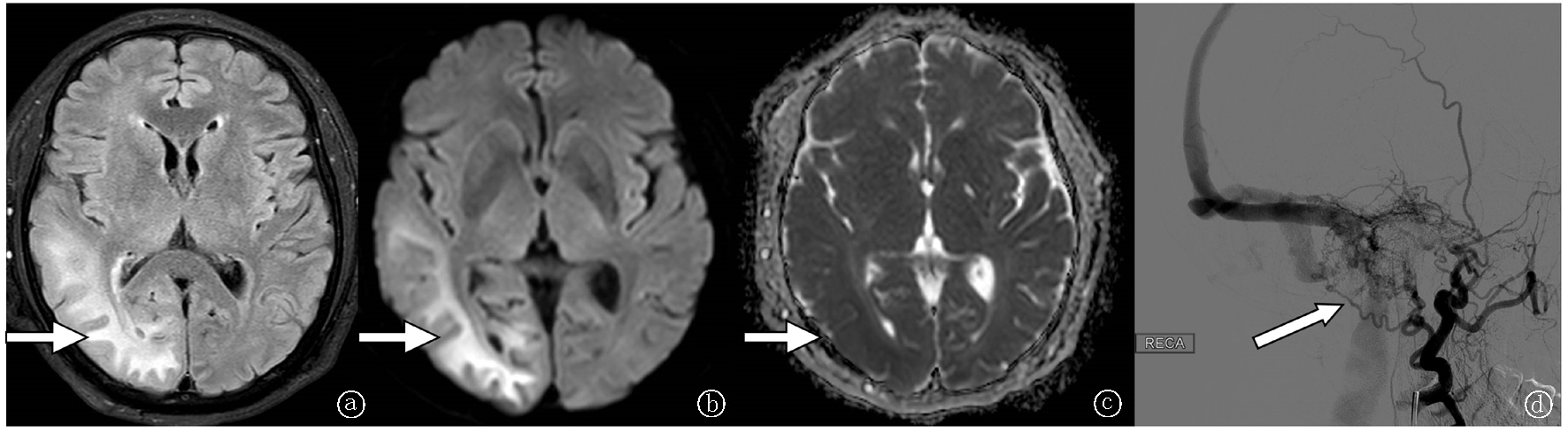
Fig.1 Case 1 a-c. Brain MRI Flair, DWI, and ADCs show abnormal signal in the right temporo-occipital lobe, suggesting the possibility of vasogenic edema; d. Right sigmoid sinus DAVF: an abnormal vascular mass is visualized in the early arterial phase, with drainage from the fistula area into the sigmoid sinus (as indicated by the arrow).
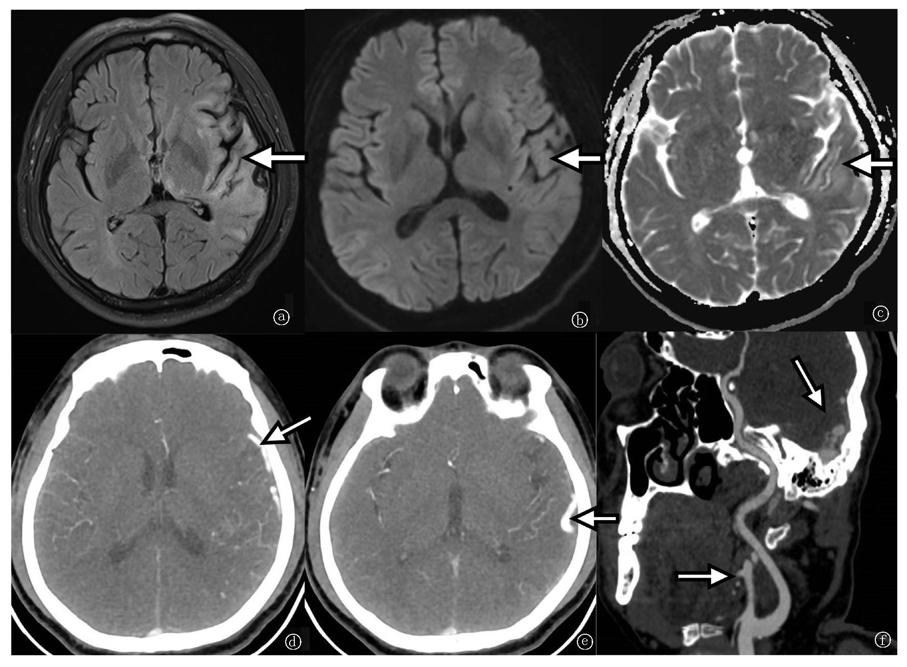
Fig.2 Case 2 a-c. Brain MRI Flair, DWI, and ADCs show cerebral infarction in the left temporal lobe; d-e. CTA shows thickened and tortuous venous vascular shadows in the left temporo-occipital lobe; f. Abnormally dilated branches of the external carotid artery and patchy abnormal enhancement in the skull base dural region, corresponding to the DAVF fistula site (as indicated by the arrow).
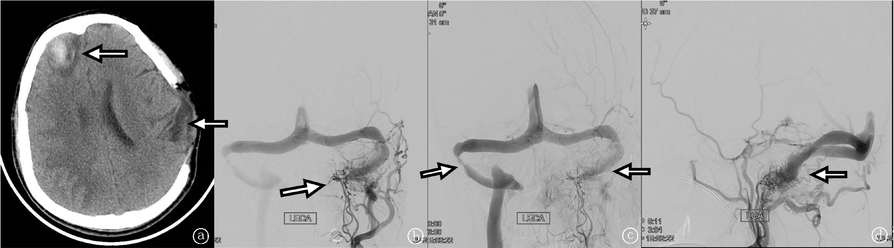
Fig.3 Case 3 a. Brain CT shows hemorrhage in the right frontal lobe and patchy low-density areas in the left temporal lobe; b-c. DSA shows left sigmoid sinus DAVF, left sigmoid sinus occlusion, and marked stenosis at the proximal segment of the right sigmoid sinus; d. Abnormal blood flow refluxes through the left petrosal sinus into the left cavernous sinus (as indicated by the arrow).
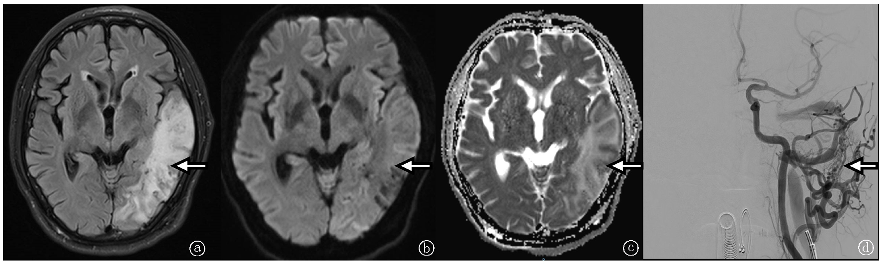
Fig.4 Case 4 a-c. Brain MRI Flair, DWI, and ADC show a subacute infarct in the left frontotemporoparietal lobe with hemorrhagic transformation; d. DAVF in the left transverse sinus and torcular region (as indicated by the arrow).
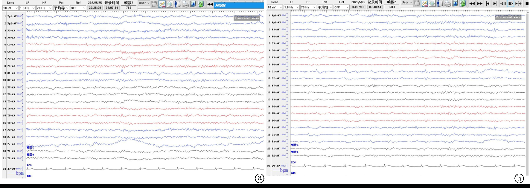
Fig.5 Long-range video EEG a. During wakefulness, periodic unilateral epileptiform discharges are seen in the left occipital region, with spike/sharp-and-slow-wave discharges in the central and parietal regions; b. During drowsiness, asynchronous scattered spikes are seen in the bilateral frontal, central, parietal, and occipital regions.
| [1] |
pmid: 5350675 |
| [2] |
|
| [3] |
pmid: 34492391 |
| [4] |
pmid: 21822156 |
| [5] |
pmid: 19190453 |
| [6] |
pmid: 7931586 |
| [7] |
|
| [8] |
pmid: 24439472 |
| [9] |
|
| [10] |
|
| [11] |
pmid: 10426146 |
| [12] |
|
| [13] |
|
| [14] |
|
| [15] |
|
| [1] | . Expert consensus on the construction and application of a 5G-based remote electroencephalography diagnosis and treatment platform for epilepsy [J]. Clinical Focus, 2026, 41(3): 264-268. |
| [2] | Task Force on the Expert Consensus for Artificial Intelligence Diagnostic Model-Assisted Electroencephalography Diagnosis of Epilepsy in the Epilepsy Subspecialty Group of the Department of Neurosurgery of the Second Hospital of Hebei Medical University. Expert consensus on deploying AI diagnostic models to support electroencephalogram-based epilepsy diagnosis [J]. Clinical Focus, 2026, 41(2): 164-168. |
| [3] | Liu Yijing, Zuo Chun, Luo Linzheng, Zhao Mingmin, Ren Bowen, Li Na. Youth stroke presenting with seizure-like onset: A case report and literature review [J]. Clinical Focus, 2026, 41(2): 155-159. |
| [4] | Wang Xiaoxiao, Hu Yanfeng, Qi Xiufeng, Zhao Junjie. Acute pulmonary embolism with epilepsy attack as the first clinical manifestation: A case report and literature review [J]. Clinical Focus, 2025, 40(6): 532-536. |
| [5] | Lin Bincheng, Lin Gangxi. Effects of etiology and preoperative course of disease on the efficacy of drug-resistant epilepsy in children [J]. Clinical Focus, 2025, 40(6): 527-531. |
| [6] | Ji Wanli, Ma Zhou, Shi Yuxi, Sun Weisheng. Spinal dural arteriovenous fistula in young: A case report and literature review [J]. Clinical Focus, 2025, 40(3): 262-265. |
| [7] | Jiao Fei, Yao Zhiping, Wang Langui. Risk factors of seizures in patients with central nervous system cystic echinococcosis [J]. Clinical Focus, 2024, 39(8): 712-715. |
| [8] | Wang Caizhen, Miao Lina, Chen Yuan, Li Shuangcheng. Effectiveness of high-frequency vagus nerve stimulation in the treatment of drug-resistant epilepsy: A meta-analysis [J]. Clinical Focus, 2024, 39(7): 593-597. |
| [9] | Zhao Shuzhen, Wang Sanping, Zhao Xiaoyun. SLC32A1 gene mutation in hereditary epilepsy with febrile convulsion addition: A case report and literature review [J]. Clinical Focus, 2023, 38(5): 451-454. |
| [10] | Ding Siqi, Liu Shihua, Zhang Chao, Zhong Ping, Cao Li. Risk factors for epilepsy after delayed post-stroke epilepsy and its clinical correlation with blood Hcy, hs-CRP and D-D [J]. Clinical Focus, 2023, 38(10): 893-897. |
| [11] | Wang Mina, Gao Weib, Hu Xiaoweia, Wu Chaob, Xu Mina. Preoperative localization and surgical outcome analysis of multifocal refractory epilepsy [J]. Clinical Focus, 2018, 33(12): 1031-1035. |
| [12] | Dong Lia, Li Xianghaib. Curative effect of oxcarbazepine on infants with partial epilepsy and immune function [J]. Clinical Focus, 2017, 32(10): 881-884. |
| Viewed | ||||||
|
Full text |
|
|||||
|
Abstract |
|
|||||