Clinical Focus ›› 2026, Vol. 41 ›› Issue (1): 38-43.doi: 10.3969/j.issn.1004-583X.2026.01.006
• Original article • Previous Articles Next Articles
Diagnostic value of CTA combined with serum TNF-α and IFN-γ for carotid plaques in ischemic stroke patients
Lu Minyana, Zhou Lia, Qi Zhiqianga, Lu Yangb( )
)
a. Department of Neurology ,Jiangsu Shengze Hospital Suzhou 215228, China b. Department of Radiology ,Jiangsu Shengze Hospital Suzhou 215228, China
-
Received:2025-10-20Online:2026-01-20Published:2026-02-02 -
Contact:Lu Yang E-mail:379940876@qq.com
CLC Number:
Cite this article
Lu Minyan, Zhou Li, Qi Zhiqiang, Lu Yang. Diagnostic value of CTA combined with serum TNF-α and IFN-γ for carotid plaques in ischemic stroke patients[J]. Clinical Focus, 2026, 41(1): 38-43.
share this article
Add to citation manager EndNote|Ris|BibTeX
URL: https://www.lchc.cn/EN/10.3969/j.issn.1004-583X.2026.01.006
| CTA检查 | DSA检查(例) | 合计 | |
|---|---|---|---|
| 有斑块 | 无斑块 | ||
| 有斑块 | 41 | 2 | 43 |
| 无斑块 | 3 | 23 | 26 |
| 合计 | 44 | 25 | 69 |
Tab.1 Consistency of CTA and DSA results
| CTA检查 | DSA检查(例) | 合计 | |
|---|---|---|---|
| 有斑块 | 无斑块 | ||
| 有斑块 | 41 | 2 | 43 |
| 无斑块 | 3 | 23 | 26 |
| 合计 | 44 | 25 | 69 |
| 组别 | 例数 | 女性 [例(%)] | 年龄 (岁) | 高血压 [例(%)] | 糖尿病 [例(%)] | TNF-α (ng/L) | IFN-γ (ng/L) |
|---|---|---|---|---|---|---|---|
| 对照组 | 66 | 20(30.3) | 67.30±12.02 | 51(77.3) | 25(37.9) | 1.07(0.57, 1.98) | 3.01(0.88, 7.80) |
| 观察组 | 69 | 19(27.5) | 63.77±11.89 | 52(75.4) | 24(34.8) | 1.78(1.24, 2.90) | 4.41(2.79, 7.57) |
| χ2/t/Z值 | 0.126 | 1.718 | 0.068 | 0.14 | -3.405 | -2.67 | |
| P值 | 0.723 | 0.088 | 0.794 | 0.708 | 0.001 | 0.008 |
Tab.2 Comparison of clinical data between two groups
| 组别 | 例数 | 女性 [例(%)] | 年龄 (岁) | 高血压 [例(%)] | 糖尿病 [例(%)] | TNF-α (ng/L) | IFN-γ (ng/L) |
|---|---|---|---|---|---|---|---|
| 对照组 | 66 | 20(30.3) | 67.30±12.02 | 51(77.3) | 25(37.9) | 1.07(0.57, 1.98) | 3.01(0.88, 7.80) |
| 观察组 | 69 | 19(27.5) | 63.77±11.89 | 52(75.4) | 24(34.8) | 1.78(1.24, 2.90) | 4.41(2.79, 7.57) |
| χ2/t/Z值 | 0.126 | 1.718 | 0.068 | 0.14 | -3.405 | -2.67 | |
| P值 | 0.723 | 0.088 | 0.794 | 0.708 | 0.001 | 0.008 |
| 组别 | 例数 | TNF-α(ng/L) | IFN-γ(ng/L) |
|---|---|---|---|
| 斑块阴性亚组 | 25 | 1.16(0.51, 1.78) | 3.56(1.24, 5.07) |
| 斑块阳性亚组 | 44 | 2.00(1.56, 4.03) | 5.09(3.87, 8.52) |
| Z值 | -3.84 | -3.223 | |
| P值 | <0.001 | 0.001 |
Tab.3 Comparison of cytokine levels between patients with and without plaques
| 组别 | 例数 | TNF-α(ng/L) | IFN-γ(ng/L) |
|---|---|---|---|
| 斑块阴性亚组 | 25 | 1.16(0.51, 1.78) | 3.56(1.24, 5.07) |
| 斑块阳性亚组 | 44 | 2.00(1.56, 4.03) | 5.09(3.87, 8.52) |
| Z值 | -3.84 | -3.223 | |
| P值 | <0.001 | 0.001 |
| 因素 | 回归系数 | 标准误 | Wald χ2值 | 值 | 值 | 95% 可信区间 | |
|---|---|---|---|---|---|---|---|
| 下限 | 上限 | ||||||
| TNF-α | 0.488 | 0.224 | 4.761 | 0.029 | 1.630 | 1.051 | 2.527 |
| IFN-γ | 0.244 | 0.11 | 4.919 | 0.027 | 1.277 | 1.029 | 1.585 |
Tab.4 Multivariate logistic regression analysis of influencing factors of carotid plaque in patients with ischemic stroke
| 因素 | 回归系数 | 标准误 | Wald χ2值 | 值 | 值 | 95% 可信区间 | |
|---|---|---|---|---|---|---|---|
| 下限 | 上限 | ||||||
| TNF-α | 0.488 | 0.224 | 4.761 | 0.029 | 1.630 | 1.051 | 2.527 |
| IFN-γ | 0.244 | 0.11 | 4.919 | 0.027 | 1.277 | 1.029 | 1.585 |
| 预测项目 | AUC | P值 | 95%可信区间 | 敏感度 | 特异度 | |
|---|---|---|---|---|---|---|
| 下限 | 上限 | |||||
| CTA | 0.926 | <0.001 | 0.850 | 1.000 | 0.932 | 0.920 |
| TNF-α | 0.780 | <0.001 | 0.662 | 0.897 | 0.909 | 0.520 |
| IFN-γ | 0.735 | 0.001 | 0.607 | 0.862 | 1.000 | 0.440 |
| 联合诊断(串联) | 0.966 | <0.001 | 0.921 | 1.000 | 0.932 | 1.000 |
Tab.5 The predictive value of a joint diagnostic model for carotid artery plaques in patients with ischemic stroke
| 预测项目 | AUC | P值 | 95%可信区间 | 敏感度 | 特异度 | |
|---|---|---|---|---|---|---|
| 下限 | 上限 | |||||
| CTA | 0.926 | <0.001 | 0.850 | 1.000 | 0.932 | 0.920 |
| TNF-α | 0.780 | <0.001 | 0.662 | 0.897 | 0.909 | 0.520 |
| IFN-γ | 0.735 | 0.001 | 0.607 | 0.862 | 1.000 | 0.440 |
| 联合诊断(串联) | 0.966 | <0.001 | 0.921 | 1.000 | 0.932 | 1.000 |
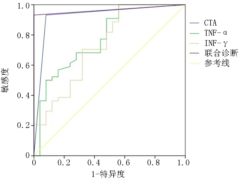
Fig.1 ROC curve of image-serum multimodal diagnostic model
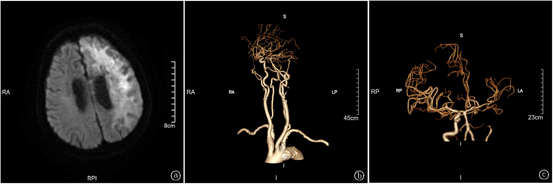
Fig.2 Typical imaging of carotid artery plaques in patients with ischemic stroke a.magnetic resonance imaging;b.CTA;c.DSA
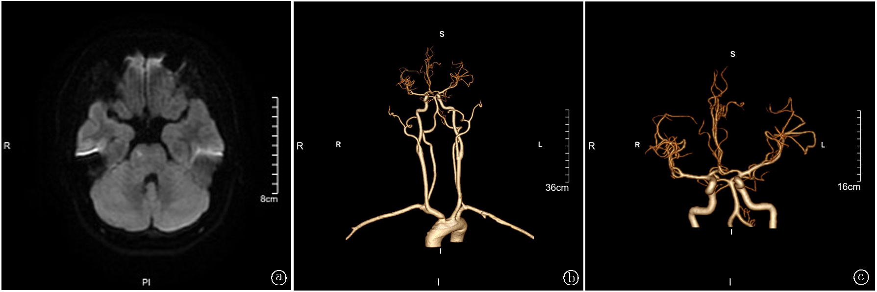
Fig.3 Typical imaging of ischemic stroke patients without carotid plaques a.magnetic resonance imaging;b.CTA;c.DSA
| [1] | Ridker PM. Anticytokine agents: Targeting interleukin signaling pathways for the treatment of atherothrombosis[J]. Circ Res, 2019, 124(3):437-450. doi:10.1161/CIRCRESAHA.118.313129. |
| [2] | Shalhoub J, Viiri LE, Cross AJ, et al. Multi-analyte profiling in human carotid atherosclerosis uncovers pro-inflammatory macrophage programming in plaques[J]. Thromb Haemost, 2016, 115(5):1064-1072. doi:10.1160/TH15-08-0650. |
| [3] | Martinez E, Martorell J, Riambau V. Review of serum biomarkers in carotid atherosclerosis[J]. J Vasc Surg, 2020, 71(1):329-341. doi: 10.1016/j.jvs.2019.04.488. |
| [4] | Ammirati E, Moroni F, Norata GD, et al. Markers of inflammation associated with plaque progression and instability in patients with carotid atherosclerosis[J]. Mediators Inflamm, 2015, 2015:718329. doi:10.1155/2015/718329. |
| [5] | Edsfeldt A, Grufman H, Asciutto G, et al. Circulating cytokines reflect the expression of pro-inflammatory cytokines in atherosclerotic plaques[J]. Atherosclerosis, 2015, 241(2):443-449. doi:10.1016/j.atherosclerosis.2015.05.019. |
| [6] | Warner JJ, Harrington RA, Sacco RL, et al. Guidelines for the early management of patients with acute ischemic stroke: 2019 update to the 2018 guidelines for the early management of acute ischemic stroke[J]. Stroke, 2019, 50(12):3331-3332. doi:10.1161/STROKEAHA.119.027708. |
| [7] | 李鑫, 柴家荣, 王彦平, 等. 三维动态增强磁共振血管壁成像在颈动脉蹼诊断中的应用[J]. 中国CT和MRI杂志, 2024, 22(3):19-21. doi:10.3969/j.issn.1672-5131.2024.03.006. |
| [8] | 戴云蛟, 艾伟平, 刘晓翠, 等. CTA联合血清MMP-9、TIMP-1对脑梗死患者颈动脉斑块诊断价值[J]. 中国CT和MRI杂志, 2024, 22(3):25-27. doi:10.3969/j.issn.1672-5131.2024.03.008. |
| [9] | GBD 2019 Stroke Collaborators. Global, regional, and national burden of stroke and its risk factors, 1990-2019: A systematic analysis for the Global Burden of Disease Study 2019[J]. Lancet Neurol, 2021, 20(10):795-820. doi: 10.1016/S1474-4422(21)00252-0. |
| [10] | Wang Y, Zhao X, Liu L, et al. Prevalence and outcomes of symptomatic intracranial large artery stenoses and occlusions in China: The Chinese Intracranial Atherosclerosis (CICAS) Study[J]. Stroke, 2014, 45(3):663-669. doi: 10.1161/STROKEAHA.113.003508. |
| [11] | 王晓民, 王国忠. 数字减影血管造影(DSA)设备的发展趋势[C]// 中华医学会医学工程学分会: 中华医学会医学工程学分会第二次医学影像设备应用技术研讨会论文集, 2001:67-69. doi:ConferenceArticle/5aa655e0c095d72220eb2237. |
| [12] | Leng X, Wong KS, Liebeskind DS. Evaluating intracranial atherosclerosis rather than intracranial stenosis[J]. Stroke, 2014, 45(2):645-651. doi:10.1161/STROKEAHA.113.002491. |
| [13] | Cloft HJ, Joseph GJ, Dion JE. Risk of cerebral angiography in patients with subarachnoid hemorrhage, cerebral aneurysm, and arteriovenous malformation: A meta-analysis[J]. Stroke, 1999, 30(2):317-320.doi: 10.1161/01.str.30.2.317. |
| [14] | Gao L, Li Z, Yuan Z, et al. Major intracranial arterial stenosis influence association between baseline blood pressure and clinical outcomes after thrombolysis in ischemic stroke patients[J]. Brain Behav, 2023, 13(6):e3022. doi: 10.1002/brb3.3022. |
| [15] | Dieleman N, van der Kolk AG, Zwanenburg JJ, et al. Imaging intracranial vessel wall pathology with magnetic resonance imaging:Current prospects and future directions[J]. Circulation, 2014, 130(2):192-201. doi:10.1161/CIRCULATIONAHA.113.006919. |
| [16] | 吕沙沙, 曹萌萌. CT血管成像与CT灌注成像对急性缺血性脑卒中的诊断价值分析[J]. 实用放射学杂志, 2020, 36(5):4.doi:10.3969/j.issn.1002-1671.2020.05.032. |
| [17] | Bäck M, Yurdagul A Jr, Tabas I, et al. Inflammation and its resolution in atherosclerosis: Mediators and therapeutic opportunities[J]. Nat Rev Cardiol, 2019, 16(7):389-406. doi: 10.1038/s41569-019-0169-2. |
| [18] | Williams JW, Huang LH, Randolph GJ. Cytokine circuits in cardiovascular disease[J]. Immunity, 2019, 50(4):941-954. doi:10.1016/j.immuni.2019.03.007. |
| [19] | Karbach S, Lagrange J, Wenzel P. Thromboinflammation and vascular dysfunction[J]. Hamostaseologie, 2019, 39(2):180-187. doi:10.1055/s-0038-1676130. |
| [20] | Engelmann B, Massberg S. Thrombosis as an intravascular effector of innate immunity[J]. Nat Rev Immunol, 2013, 13(1):34-45. doi:10.1038/nri3345. |
| [21] | D'Alessandro E, Becker C, Bergmeier W, et al. Thrombo-inflammation in cardiovascular disease: An Expert consensus document from the third maastricht consensus conference on thrombosis[J]. Thromb Haemost, 2020, 120(4):538-564. doi:10.1055/s-0040-1708035. |
| [22] | Pfeffer K. Biological functions of tumor necrosis factor cytokines and their receptors[J]. Cytokine Growth Factor Rev, 2003, 14(3-4):185-191. doi: 10.1016/S1359-6101(03)00022-4. |
| [23] | Ma X, Yao H, Yang Y, et al. miR-195 suppresses abdominal aortic aneurysm through the TNF-α/NF-κB and VEGF/PI3K/Akt pathway[J]. Int J Mol Med, 2018, 41(4):2350-2358. doi: 10.3892/ijmm.2018.3426. |
| [24] | Batra R, Suh MK, Carson JS, et al. IL-1β (Interleukin-1β) and TNF-α (Tumor Necrosis Factor-α) impact abdominal aortic aneurysm formation by differential effects on macrophage polarization[J]. Arterioscler Thromb Vasc Biol, 2017, 38(2):457-463. doi: 10.1161/ATVBAHA.117.310333. |
| [25] | Liu P, Zhang C, Chen J, et al. Combinational therapy of interferon-α and chemotherapy normalizes tumor vasculature by regulating pericytes including the novel marker RGS5 in melanoma.[J]. J Immunother, 2011, 34(3):320-326. doi: 10.1097/CJI.0b013e318213cd12. |
| [26] | Schrimpf C, Koppen T, Duffield JS, et al. TIMP3 is regulated by pericytes upon shear stress detection leading to a modified endothelial cell response[J]. Eur J Vasc Endovasc Surg, 2017, 54(4):524-533. doi:10.1016/j.jvs.2017.08.037. |
| Viewed | ||||||
|
Full text |
|
|||||
|
Abstract |
|
|||||