临床荟萃 ›› 2026, Vol. 41 ›› Issue (8): 705-710.doi: 10.3969/j.issn.1004-583X.2026.08.006

• 论著 • 上一篇    下一篇

右向左分流与脑白质病变严重程度的相关性

郭一鸣, 赵海娜, 陈丹妮, 陈蓓蕾, 李晓波, 姜超()   

  1. 苏北人民医院 神经内科, 江苏 扬州 225009
  • 收稿日期:2026-07-14 出版日期:2026-08-20 发布日期:2026-08-25
  • 通讯作者: 姜超 E-mail:605201948@qq.com

Association between right-to-left shunt and the severity of white matter lesions

Guo Yiming, Zhao Haina, Chen Danni, Chen Beilei, Li Xiaobo, Jiang Chao()   

  1. Department of Neurology, Northern Jiangsu People’s Hospital, Yangzhou 225009, China
  • Received:2026-07-14 Online:2026-08-20 Published:2026-08-25
  • Contact: Jiang Chao E-mail:605201948@qq.com

摘要:

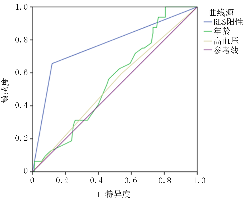
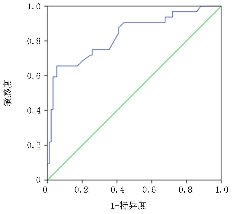
目的 探讨右向左分流(right-to-left shunt, RLS)与脑白质病变(white matter lesions, WML)严重程度之间的相关性。方法 连续纳入自2022年1月-2025年7月于苏北人民医院神经内科住院并完善经颅多普勒超声发泡试验的患者。收集基线信息及临床特征等相关资料,所有入组患者均进行经颅多普勒超声发泡试验及头颅磁共振检查。WML的评定为:侧脑室旁或深部脑白质区域T2加权像及FLAIR成像高信号。WML使用Fazekas量表进行评分。轻度WML定义为0~3分,重度WML定义为4~6分。RLS定义为:经颅多普勒超声发泡试验提示单通道双深度模式下单侧大脑中动脉监测过程中至少有一个及以上微栓子信号。 结果 本研究共计纳入125例完善经颅多普勒超声发泡试验的住院患者,其中轻度WML组93例(74.4%),重度WML组32例(25.6%)。RLS阳性32例(25.6%)。采用Mann-Whitney U检验进行单因素分析发现,与轻度WML组相比,重度WML组RLS阳性患者占比更多(P<0.01);中性粒细胞计数水平显著升高(P=0.003);中性粒细胞/淋巴细胞比值(neutrophil-to-lymphocyte ratio,NLR)水平显著升高(P=0.004)。简明精神状态检查表评分显著降低(P<0.01)。以RLS阳性、NLR水平、年龄、高血压、糖尿病作为自变量进行二元Logistic回归分析结果显示,RLS阳性(OR=39.290, 95%CI:9.54~161.74,P<0.01)、年龄(OR=1.090, 95%CI:1.02-1.16,P=0.011)、高血压(OR=3.749, 95%CI:1.02~13.84,P=0.047)是WML严重程度的独立危险因素。单指标ROC曲线分析显示,RLS阳性预测WML严重程度的曲线下面积(AUC)为0.769(95%CI:0.663~0.875,P<0.01),单独预测效能良好,而年龄(AUC=0.557)、高血压(AUC=0.528)单独预测效能较低。将RLS阳性、年龄、高血压纳入二元 Logistic回归构建联合预测模型,根据其预测概率绘制ROC曲线,结果显示AUC=0.831(95%CI:0.743~0.920,P<0.01),最佳截断值为0.431,敏感度和特异度分别为0.656和0.946,提示多指标联合模型对WML严重程度具有较好的预测价值。结论 WML严重程度可能与是否合并RLS存在一定的相关性。

关键词: 脑白质病, 右向左分流, 经颅多普勒超声发泡试验

Abstract:

Objective To investigate the association between right-to-left shunt (RLS) and the severity of white matter lesions (WML). Methods Patients hospitalized in the Department of Neurology of Northern Jiangsu People’s Hospital from January 2022 to July 2025 who underwent contrast-enhanced transcranial doppler were consecutively enrolled. Baseline information and clinical characteristics were collected. All enrolled patients underwent contrast-enhanced transcranial doppler and brain magnetic resonance imaging. WML was defined as hyperintense signals on T2-weighted and fluid-attenuated inversion recovery (FLAIR) images in the periventricular or deep white matter regions. WML severity was assessed using the Fazekas scale. Mild WML was defined as 0-3 points, and severe WML as 4-6 points. RLS was defined as the presence of at least one microembolic signal in the unilateral middle cerebral artery monitored under the single-channel double-depth mode during contrast-enhanced transcranial doppler. Results A total of 125 hospitalized patients who completed contrast-enhanced transcranial doppler were included, including 93 patients in the mild WML group (74.4%) and 32 patients in the severe WML group (25.6%). RLS was positive in 32 patients (25.6%). Univariate analysis using the Mann-Whitney U test showed that, compared with the mild WML group, the severe WML group had a higher proportion of RLS-positive patients (P<0.01), significantly higher neutrophil count (P=0.003), and significantly higher neutrophil-to-lymphocyte ratio (NLR)(P=0.004). The Mini-Mental State Examination score was significantly lower in the severe WML group (P<0.01). Binary logistic regression with RLS positivity, NLR, age, hypertension, and diabetes as independent variables showed that RLS positivity (OR=39.290, 95%CI: 9.54-161.74, P<0.01), age (OR=1.090, 95%CI: 1.02-1.16, P=0.011), and hypertension (OR=3.749, 95%CI: 1.02-13.84, P=0.047) were independent risk factors for WML severity. Single-factor ROC curve analysis showed that the area under the curve (AUC) of RLS positivity for predicting WML severity was 0.76 (95%CI: 0.663-0.875, P<0.01), indicating good predictive performance, whereas age (AUC=0.557) and hypertension (AUC=0.528) showed relatively poor independent predictive performance. A combined predictive model was constructed by incorporating RLS positivity, age, and hypertension into binary logistic regression, and an ROC curve was generated based on the predicted probability. The results showed an AUC of 0.831(95%CI: 0.743-0.920, P<0.01), with an optimal cutoff value of 0.431, sensitivity of 0.656, and specificity of 0.946, suggesting that the multi-indicator combined model has good predictive value for WML severity.Conclusion The severity of WML may be associated with the presence of RLS.

Key words: leukoencephalopathies, right-to-left shunt, contrast-enhanced transcranial doppler

中图分类号: